2WWR image
Deposition Date 2009-10-26
Release Date 2010-10-13
Last Version Date 2023-12-20
Entry Detail
PDB ID:
2WWR
Keywords:
Title:
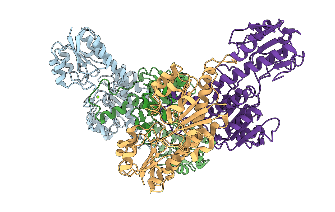
Crystal Structure of Human Glyoxylate Reductase Hydroxypyruvate Reductase
Biological Source:
Source Organism:
HOMO SAPIENS (Taxon ID: 9606)
Host Organism:
Method Details:
Experimental Method:
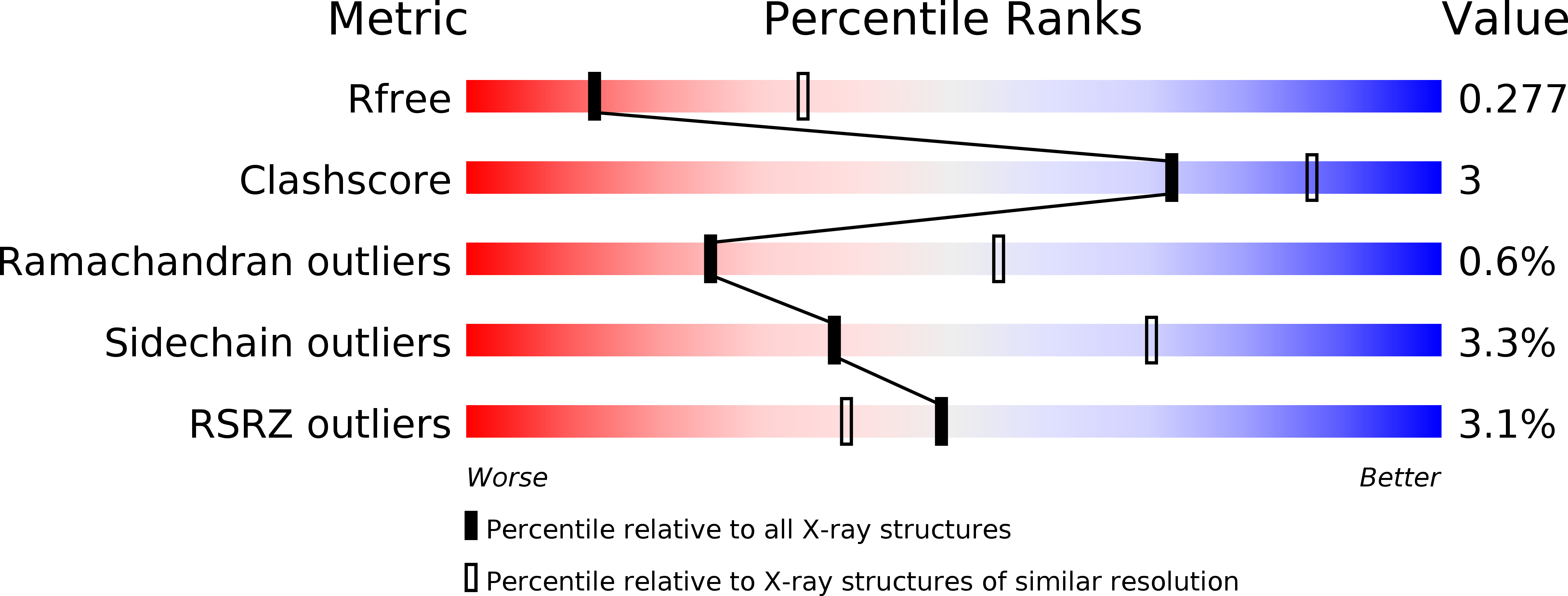
Resolution:
2.82 Å
R-Value Free:
0.27
R-Value Work:
0.21
R-Value Observed:
0.22
Space Group:
P 21 21 21
Macromolecular Entities
Polymer Type:polypeptide(L)
Molecule:GLYOXYLATE REDUCTASE/HYDROXYPYRUVATE REDUCTASE
Gene (Uniprot):GRHPR
Chain IDs:A, B, C, D
Chain Length:330
Number of Molecules:4
Biological Source:HOMO SAPIENS
Ligand Molecules
Primary Citation
Structural Basis of Substrate Specificity in Human Glyoxylate Reductase/Hydroxypyruvate Reductase.
J.Mol.Biol. 360 178 ? (2006)
PMID: 16756993 DOI: 10.1016/J.JMB.2006.05.018

Abstact

Human glyoxylate reductase/hydroxypyruvate reductase (GRHPR) is a D-2-hydroxy-acid dehydrogenase that plays a critical role in the removal of the metabolic by-product glyoxylate from within the liver. Deficiency of this enzyme is the underlying cause of primary hyperoxaluria type 2 (PH2) and leads to increased urinary oxalate levels, formation of kidney stones and renal failure. Here we describe the crystal structure of human GRHPR at 2.2 A resolution. There are four copies of GRHPR in the crystallographic asymmetric unit: in each homodimer, one subunit forms a ternary (enzyme+NADPH+reduced substrate) complex, and the other a binary (enzyme+NADPH) form. The spatial arrangement of the two enzyme domains is the same in binary and ternary forms. This first crystal structure of a true ternary complex of an enzyme from this family demonstrates the relationship of substrate and catalytic residues within the active site, confirming earlier proposals of the mode of substrate binding, stereospecificity and likely catalytic mechanism for these enzymes. GRHPR has an unusual substrate specificity, preferring glyoxylate and hydroxypyruvate, but not pyruvate. A tryptophan residue (Trp141) from the neighbouring subunit of the dimer is projected into the active site region and appears to contribute to the selectivity for hydroxypyruvate. This first crystal structure of a human GRHPR enzyme also explains the deleterious effects of naturally occurring missense mutations of this enzyme that lead to PH2.

Legend

Protein

Chemical

Disease

Primary Citation of related structures