2WVQ image
Deposition Date 2009-10-19
Release Date 2010-07-28
Last Version Date 2023-12-20
Entry Detail
PDB ID:
2WVQ
Title:
Structure of the HET-s N-terminal domain. Mutant D23A, P33H
Biological Source:
Source Organism:
Host Organism:
Method Details:
Experimental Method:
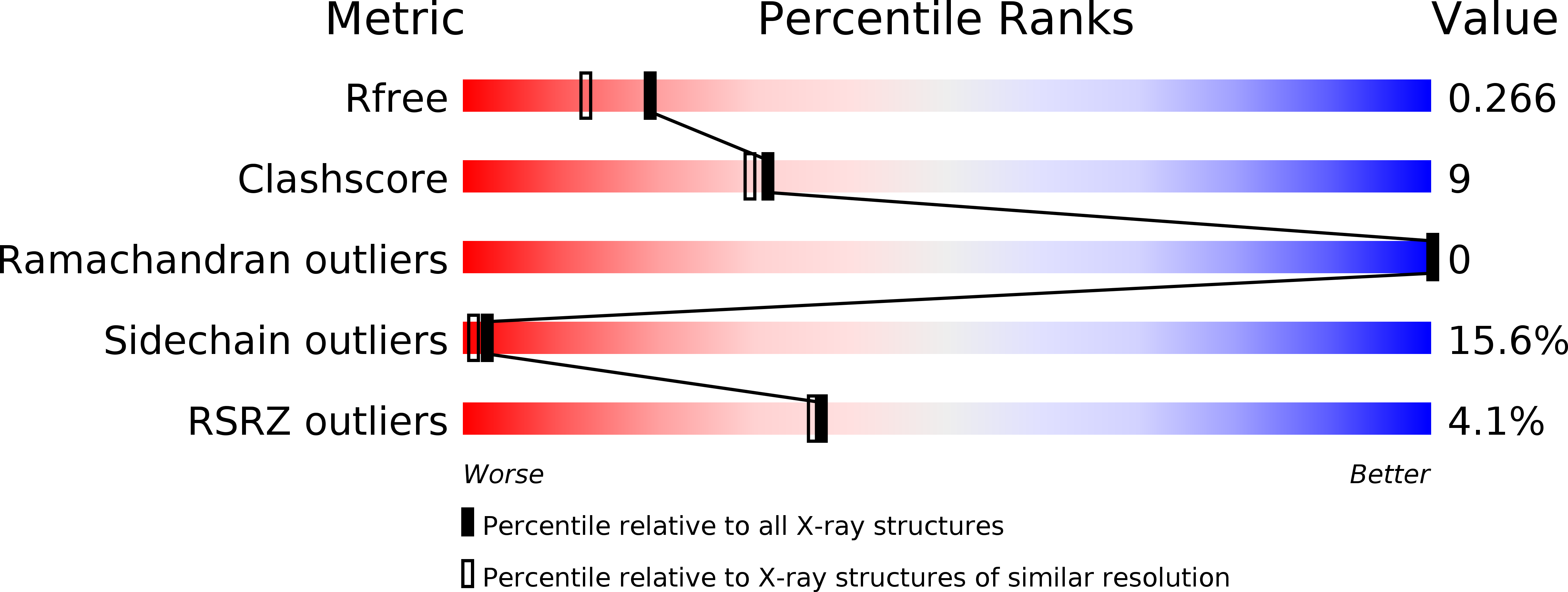
Resolution:
2.00 Å
R-Value Free:
0.26
R-Value Work:
0.22
R-Value Observed:
0.22
Space Group:
P 1 21 1
Macromolecular Entities
Structures with similar UniProt ID
Protein Blast
Polymer Type:polypeptide(L)
Molecule:SMALL S PROTEIN
Gene (Uniprot):het-s
Mutagens:YES
Chain IDs:A, B
Chain Length:225
Number of Molecules:2
Biological Source:PODOSPORA ANSERINA
Primary Citation
The mechanism of prion inhibition by HET-S.
Mol. Cell 38 889 899 (2010)
PMID: 20620958 DOI: 10.1016/j.molcel.2010.05.019

Abstact

HET-S (97% identical to HET-s) has an N-terminal globular domain that exerts a prion-inhibitory effect in cis on its own prion-forming domain (PFD) and in trans on HET-s prion propagation. We show that HET-S fails to form fibrils in vitro and that it inhibits HET-s PFD fibrillization in trans. In vivo analyses indicate that beta-structuring of the HET-S PFD is required for HET-S activity. The crystal structures of the globular domains of HET-s and HET-S are highly similar, comprising a helical fold, while NMR-based characterizations revealed no differences in the conformations of the PFDs. We conclude that prion inhibition is not encoded by structure but rather in stability and oligomerization properties: when HET-S forms a prion seed or is incorporated into a HET-s fibril via its PFD, the beta-structuring in this domain induces a change in its globular domain, generating a molecular species that is incompetent for fibril growth.

Legend

Protein

Chemical

Disease

Primary Citation of related structures