Deposition Date
2009-10-15
Release Date
2009-12-01
Last Version Date
2023-12-20
Entry Detail
PDB ID:
2WV9
Keywords:
Title:
Crystal Structure of the NS3 protease-helicase from Murray Valley encephalitis virus
Biological Source:
Source Organism(s):
MURRAY VALLEY ENCEPHALITIS VIRUS (Taxon ID: 301478)
Expression System(s):
Method Details:
Experimental Method:
Resolution:
2.75 Å
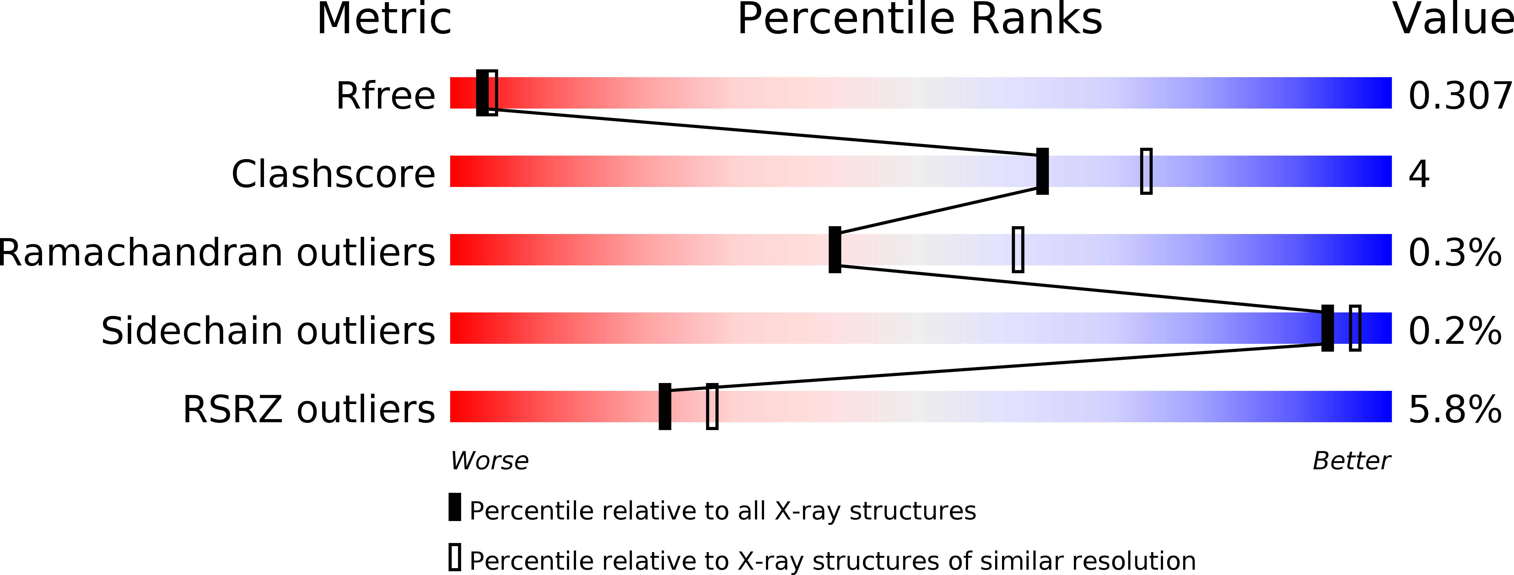
R-Value Free:
0.30
R-Value Work:
0.26
R-Value Observed:
0.26
Space Group:
P 1 21 1