2WUR image
Deposition Date 2009-10-07
Release Date 2010-05-12
Last Version Date 2024-11-06
Entry Detail
PDB ID:
2WUR
Title:
Atomic resolution structure of GFP measured on a rotating anode
Biological Source:
Source Organism:
Host Organism:
Method Details:
Experimental Method:
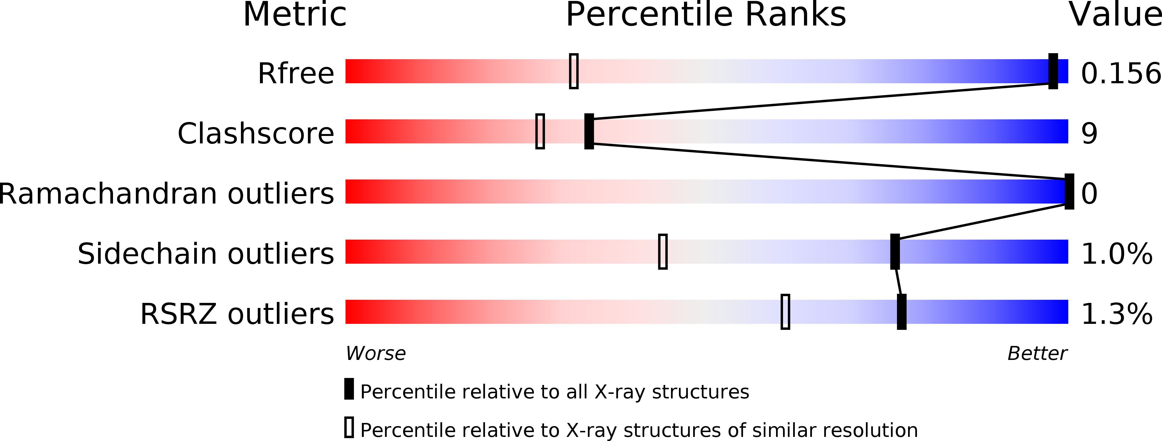
Resolution:
0.90 Å
R-Value Free:
0.17
Space Group:
P 21 21 21
Macromolecular Entities
Polymer Type:polypeptide(L)
Molecule:GREEN FLUORESCENT PROTEIN
Gene (Uniprot):GFP
Mutations:YES
Chain IDs:A
Chain Length:238
Number of Molecules:1
Biological Source:AEQUOREA VICTORIA
Modified Residue
Compound ID Chain ID Parent Comp ID Details 2D Image
GYS A SER ?
Primary Citation
Visualizing Proton Antenna in a High-Resolution Green Fluorescent Protein Structure.
J.Am.Chem.Soc. 132 11093 ? (2010)
PMID: 20698675 DOI: 10.1021/JA1010652

Abstact

"Proton-collecting antenna" are conjectured to consist of several carboxylates within hydrogen-bond (HB) networks on the surface of proteins, which funnel protons to the orifice of an internal proton wire leading to the protein's active site. Yet such constructions were never directly visualized. Here we report an X-ray structure of green fluorescent protein (GFP) of the highest resolution to date (0.9 A). It allows the identification of some pivotal hydrogen atoms pertinent to uncertainties concerning the protonation state of the chromophore. Applying a computer algorithm for mapping proton wires in proteins reveals the previously observed "active site wire" connecting Glu222 with the surface carboxylate Glu5. In addition, it is now possible to identify what appears to be a proton-collecting apparatus of GFP. It consists of a negative surface patch containing carboxylates, threonines, and water molecules, connected by a HB network to Glu5. Furthermore, we detect exit points via Asn146 and His148 to a hydrophobic surface region. The more extensive HB network of the present structure, as compared with earlier GFP structures, is not accidental. A systematic investigation of over 100 mutants shows a clear correlation between the observed water content of GFP X-ray structures and their resolution. With increasing water content, the proton wires become progressively larger. These findings corroborate the scenario in which the photodissociated proton from wild-type GFP can leak outside, whereafter another proton is recruited via the proton-collecting apparatus reported herein.

Legend

Protein

Chemical

Disease