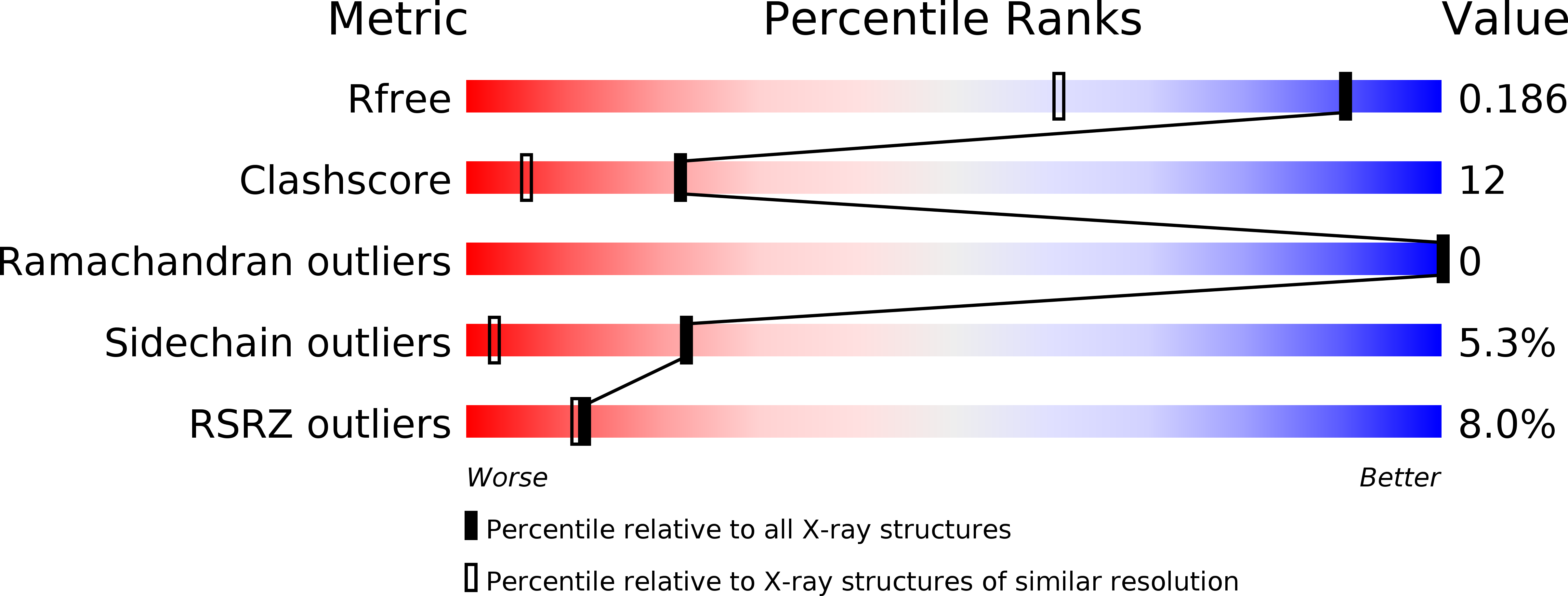
2WUJ image
Deposition Date 2009-10-06
Release Date 2010-06-09
Last Version Date 2024-05-08
Entry Detail
PDB ID:
2WUJ
Keywords:
Title:
DivIVA N-terminal domain
Biological Source:
Source Organism:
Host Organism:
Method Details:
Experimental Method:
Resolution:
1.40 Å
R-Value Free:
0.22
R-Value Work:
0.18
R-Value Observed:
0.18
Space Group:
P 21 21 21
Macromolecular Entities
Structures with similar UniProt ID
Protein Blast
Polymer Type:polypeptide(L)
Molecule:SEPTUM SITE-DETERMINING PROTEIN DIVIVA
Gene (Uniprot):divIVA
Chain IDs:A, B
Chain Length:57
Number of Molecules:2
Biological Source:BACILLUS SUBTILIS
Primary Citation
Features Critical for Membrane Binding Revealed by Diviva Crystal Structure.
Embo J. 29 1988 ? (2010)
PMID: 20502438 DOI: 10.1038/EMBOJ.2010.99

Abstact

DivIVA is a conserved protein in Gram-positive bacteria that localizes at the poles and division sites, presumably through direct sensing of membrane curvature. DivIVA functions as a scaffold and is vital for septum site selection during vegetative growth and chromosome anchoring during sporulation. DivIVA deletion causes filamentous growth in Bacillus subtilis, whereas overexpression causes hyphal branching in Streptomyces coelicolor. We have determined the crystal structure of the N-terminal (Nt) domain of DivIVA, and show that it forms a parallel coiled-coil. It is capped with two unique crossed and intertwined loops, exposing hydrophobic and positively charged residues that we show here are essential for membrane binding. An intragenic suppressor introducing a positive charge restores membrane binding after mutating the hydrophobic residues. We propose that the hydrophobic residues insert into the membrane and that the positively charged residues bind to the membrane surface. A low-resolution crystal structure of the C-terminal (Ct) domain displays a curved tetramer made from two parallel coiled-coils. The Nt and Ct parts were then merged into a model of the full length, 30 nm long DivIVA protein.

Legend

Protein

Chemical

Disease

Primary Citation of related structures