Deposition Date
2009-10-01
Release Date
2009-10-27
Last Version Date
2023-12-20
Entry Detail
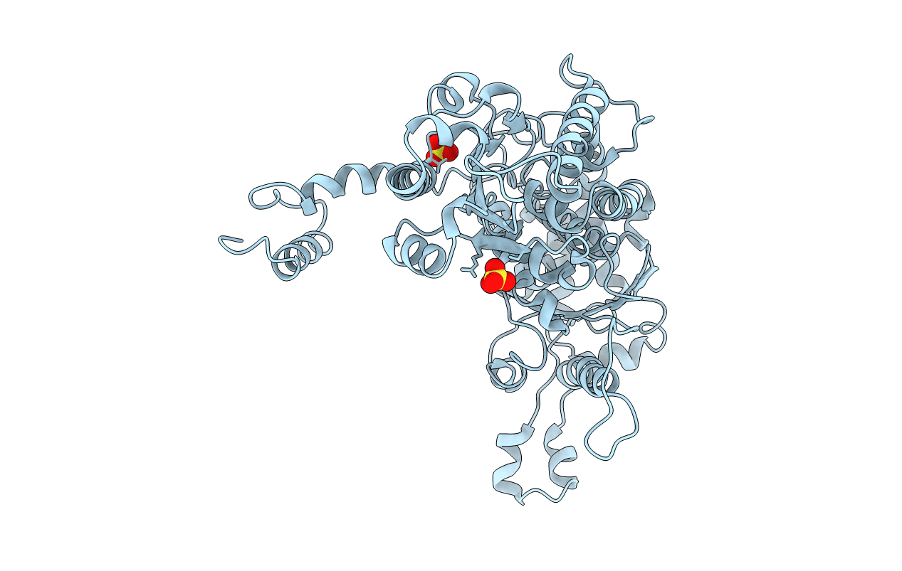
PDB ID:
2WU8
Keywords:
Title:
Structural studies of phosphoglucose isomerase from Mycobacterium tuberculosis H37Rv
Biological Source:
Source Organism(s):
MYCOBACTERIUM TUBERCULOSIS (Taxon ID: 83332)
Expression System(s):
Method Details:
Experimental Method:
Resolution:
2.25 Å
R-Value Free:
0.22
R-Value Work:
0.18
R-Value Observed:
0.18
Space Group:
I 21 21 21