
Deposition Date
2009-09-16
Release Date
2010-04-21
Last Version Date
2024-05-08
Entry Detail
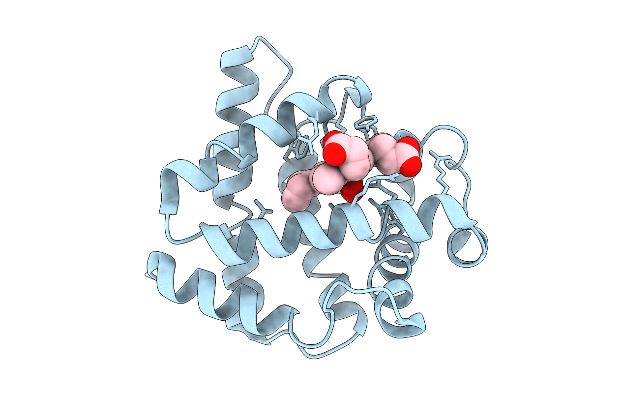
PDB ID:
2WTG
Keywords:
Title:
High resolution 3D structure of C.elegans globin-like protein GLB-1
Biological Source:
Source Organism(s):
CAENORHABDITIS ELEGANS (Taxon ID: 6239)
Expression System(s):
Method Details:
Experimental Method:
Resolution:
1.50 Å
R-Value Free:
0.20
R-Value Work:
0.16
R-Value Observed:
0.16
Space Group:
P 43 21 2
