2WSS image
Deposition Date 2009-09-09
Release Date 2009-11-17
Last Version Date 2024-11-13
Entry Detail
PDB ID:
2WSS
Keywords:
Title:
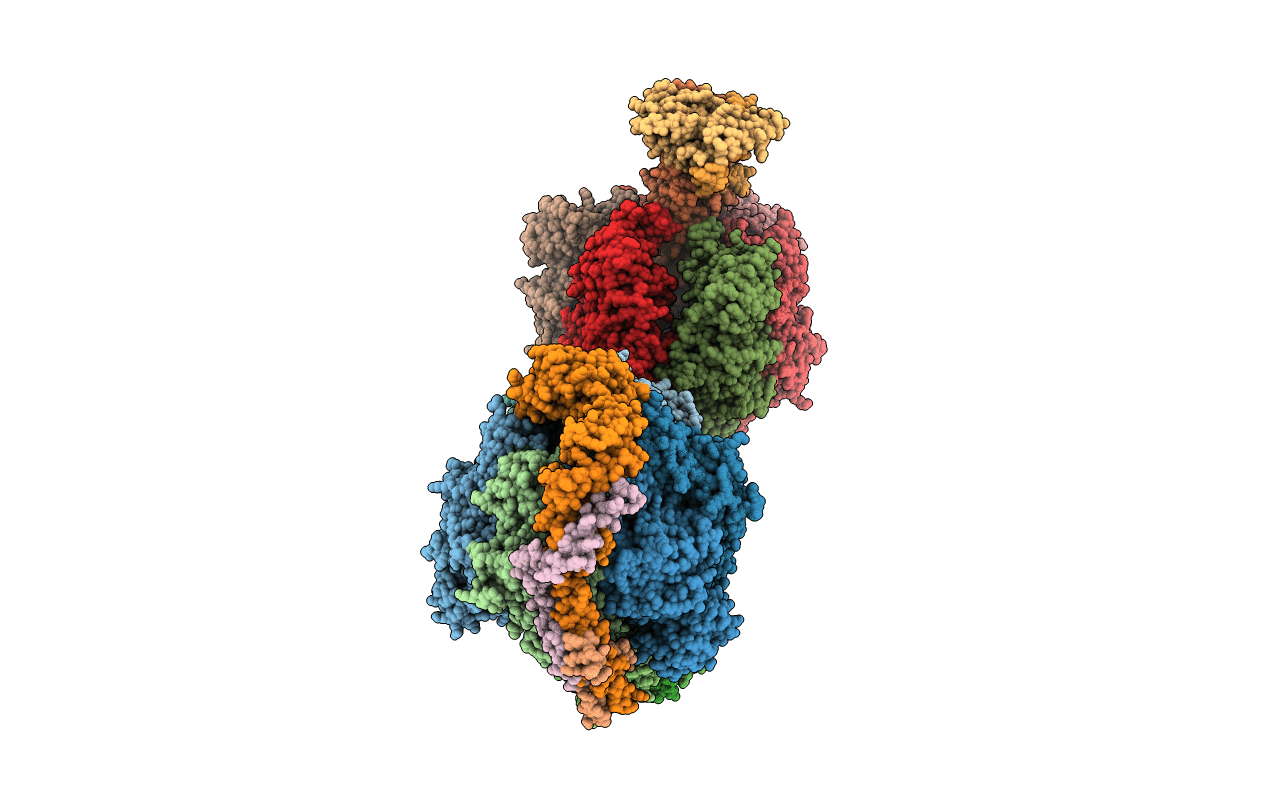
The structure of the membrane extrinsic region of bovine ATP synthase
Biological Source:
Source Organism:
BOS TAURUS (Taxon ID: 9913)
Host Organism:
Method Details:
Experimental Method:
Resolution:
3.20 Å
R-Value Free:
0.27
R-Value Work:
0.21
R-Value Observed:
0.22
Space Group:
P 21 21 21
Macromolecular Entities
Structures with similar UniProt ID
Protein Blast
Polymer Type:polypeptide(L)
Molecule:ATP SYNTHASE SUBUNIT ALPHA, MITOCHONDRIAL
Gene (Uniprot):ATP5F1A
Chain IDs:A, B, C, J, K, L
Chain Length:510
Number of Molecules:6
Biological Source:BOS TAURUS
Structures with similar UniProt ID
Protein Blast
Polymer Type:polypeptide(L)
Molecule:ATP SYNTHASE SUBUNIT BETA, MITOCHONDRIAL
Gene (Uniprot):ATP5F1B
Chain IDs:D, E, F, M, N, O
Chain Length:482
Number of Molecules:6
Biological Source:BOS TAURUS
Structures with similar UniProt ID
Protein Blast
Polymer Type:polypeptide(L)
Molecule:ATP SYNTHASE SUBUNIT GAMMA, MITOCHONDRIAL
Gene (Uniprot):ATP5F1C
Chain IDs:G, P
Chain Length:272
Number of Molecules:2
Biological Source:BOS TAURUS
Structures with similar UniProt ID
Protein Blast
Polymer Type:polypeptide(L)
Molecule:ATP SYNTHASE SUBUNIT DELTA, MITOCHONDRIAL
Gene (Uniprot):ATP5F1D
Chain IDs:H, Q
Chain Length:146
Number of Molecules:2
Biological Source:BOS TAURUS
Structures with similar UniProt ID
Protein Blast
Polymer Type:polypeptide(L)
Molecule:ATP SYNTHASE SUBUNIT EPSILON, MITOCHONDRIAL
Gene (Uniprot):ATP5F1E
Chain IDs:I, R
Chain Length:50
Number of Molecules:2
Biological Source:BOS TAURUS
Structures with similar UniProt ID
Protein Blast
Polymer Type:polypeptide(L)
Molecule:ATP SYNTHASE SUBUNIT O, MITOCHONDRIAL
Gene (Uniprot):ATP5PO
Chain IDs:S, W
Chain Length:190
Number of Molecules:2
Biological Source:BOS TAURUS
Structures with similar UniProt ID
Protein Blast
Polymer Type:polypeptide(L)
Molecule:ATP SYNTHASE SUBUNIT B, MITOCHONDRIAL
Gene (Uniprot):ATP5PB
Chain IDs:T, X
Chain Length:116
Number of Molecules:2
Biological Source:BOS TAURUS
Structures with similar UniProt ID
Protein Blast
Polymer Type:polypeptide(L)
Molecule:ATP SYNTHASE SUBUNIT D, MITOCHONDRIAL
Gene (Uniprot):ATP5PD
Chain IDs:U
Chain Length:118
Number of Molecules:1
Biological Source:BOS TAURUS
Structures with similar UniProt ID
Protein Blast
Polymer Type:polypeptide(L)
Molecule:ATP SYNTHASE-COUPLING FACTOR 6, MITOCHONDRIAL
Gene (Uniprot):ATP5PF
Chain IDs:V, Y (auth: Z)
Chain Length:76
Number of Molecules:2
Biological Source:BOS TAURUS
Modified Residue
Compound ID Chain ID Parent Comp ID Details 2D Image
MSE S MET SELENOMETHIONINE
Primary Citation
The Structure of the Membrane Extrinsic Region of Bovine ATP Synthase
Proc.Natl.Acad.Sci.USA 106 21597 ? (2009)
PMID: 19995987 DOI: 10.1073/PNAS.0910365106

Abstact

The structure of the complex between bovine mitochondrial F(1)-ATPase and a stator subcomplex has been determined at a resolution of 3.2 A. The resolved region of the stator contains residues 122-207 of subunit b; residues 5-25 and 35-57 of F(6); 3 segments of subunit d from residues 30-40, 65-74, and 85-91; and residues 1-146 and 169-189 of the oligomycin sensitivity conferral protein (OSCP). The stator subcomplex represents its membrane distal part, and its structure has been augmented with an earlier structure of a subcomplex containing residues 79-183, 3-123, and 5-70 of subunits b, d, and F(6), respectively, which extends to the surface of the inner membrane of the mitochondrion. The N-terminal domain of the OSCP links the stator with F(1)-ATPase via alpha-helical interactions with the N-terminal region of subunit alpha(E). Its C-terminal domain makes extensive helix-helix interactions with the C-terminal alpha-helix of subunit b from residues 190-207. Subunit b extends as a continuous 160-A long alpha-helix from residue 188 back to residue 79 near to the surface of the inner mitochondrial membrane. This helix appears to be stiffened by other alpha-helices in subunits d and F(6), but the structure can bend inward toward the F(1) domain around residue 146 of subunit b. The linker region between the 2 domains of the OSCP also appears to be flexible, enabling the stator to adjust its shape as it passes over the changing profile of the F(1) domain during a catalytic cycle. The structure of the membrane extrinsic part of bovine ATP synthase is now complete.

Legend

Protein

Chemical

Disease