
Deposition Date
2009-09-04
Release Date
2010-09-29
Last Version Date
2023-12-20
Entry Detail
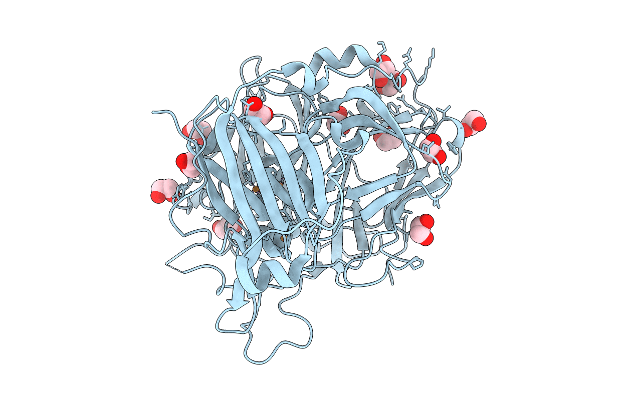
PDB ID:
2WSD
Keywords:
Title:
Proximal mutations at the type 1 Cu site of CotA-laccase: I494A mutant
Biological Source:
Source Organism(s):
BACILLUS SUBTILIS (Taxon ID: 1423)
Expression System(s):
Method Details:
Experimental Method:
Resolution:
1.60 Å
R-Value Free:
0.19
R-Value Work:
0.17
R-Value Observed:
0.17
Space Group:
P 31 2 1
