Deposition Date
1998-08-04
Release Date
1999-08-04
Last Version Date
2024-11-13
Entry Detail
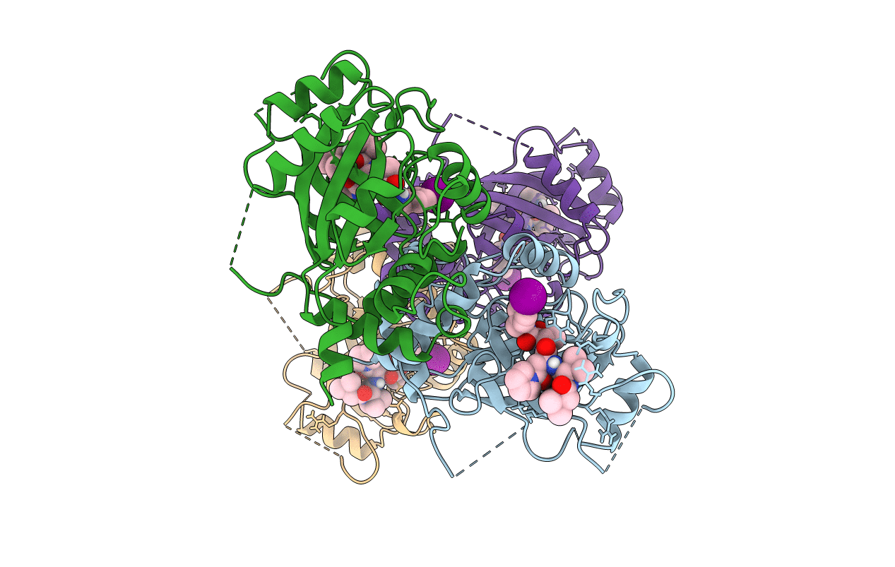
Biological Source:
Source Organism(s):
Human herpesvirus 5 (Taxon ID: 10359)
Expression System(s):
Method Details:
Experimental Method:
Resolution:
2.70 Å
R-Value Free:
0.33
R-Value Work:
0.22
R-Value Observed:
0.22
Space Group:
P 21 2 21