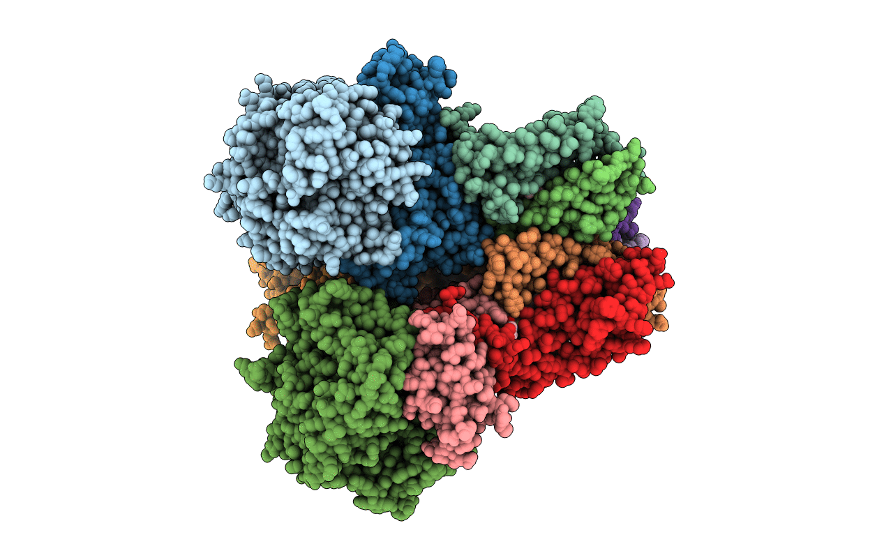
2WP9 image
Entry Detail
PDB ID:
2WP9
Keywords:
Title:
Crystal structure of the E. coli succinate:quinone oxidoreductase (SQR) SdhB His207Thr mutant
Biological Source:
Source Organism:
Host Organism:
PDB Version:
Deposition Date:
2009-08-03
Release Date:
2010-08-25
Method Details:
Experimental Method:
Resolution:
2.70 Å
R-Value Free:
0.22
R-Value Work:
0.19
R-Value Observed:
0.19
Space Group:
P 21 21 21
Macromolecular Entities
Polymer Type:polypeptide(L)
Description:SUCCINATE DEHYDROGENASE FLAVOPROTEIN SUBUNIT
Chain IDs:A, E, I
Chain Length:588
Number of Molecules:3
Biological Source:ESCHERICHIA COLI
Polymer Type:polypeptide(L)
Description:SUCCINATE DEHYDROGENASE IRON-SULFUR SUBUNIT
Mutations:YES
Chain IDs:B, F, J
Chain Length:238
Number of Molecules:3
Biological Source:ESCHERICHIA COLI
Polymer Type:polypeptide(L)
Description:SUCCINATE DEHYDROGENASE CYTOCHROME B556 SUBUNIT
Chain IDs:C, G, K
Chain Length:129
Number of Molecules:3
Biological Source:ESCHERICHIA COLI
Polymer Type:polypeptide(L)
Description:SUCCINATE DEHYDROGENASE HYDROPHOBIC MEMBRANE ANCHOR SUBUNIT
Chain IDs:D, H, L
Chain Length:115
Number of Molecules:3
Biological Source:ESCHERICHIA COLI
Primary Citation
Perturbation of the quinone-binding site of complex II alters the electronic properties of the proximal [3Fe-4S] iron-sulfur cluster.
J. Biol. Chem. 286 12756 12765 (2011)
PMID: 21310949 DOI: 10.1074/jbc.M110.209874

Abstact

Succinate-ubiquinone oxidoreductase (SQR) and menaquinol-fumarate oxidoreductase (QFR) from Escherichia coli are members of the complex II family of enzymes. SQR and QFR catalyze similar reactions with quinones; however, SQR preferentially reacts with higher potential ubiquinones, and QFR preferentially reacts with lower potential naphthoquinones. Both enzymes have a single functional quinone-binding site proximal to a [3Fe-4S] iron-sulfur cluster. A difference between SQR and QFR is that the redox potential of the [3Fe-4S] cluster in SQR is 140 mV higher than that found in QFR. This may reflect the character of the different quinones with which the two enzymes preferentially react. To investigate how the environment around the [3Fe-4S] cluster affects its redox properties and catalysis with quinones, a conserved amino acid proximal to the cluster was mutated in both enzymes. It was found that substitution of SdhB His-207 by threonine (as found in QFR) resulted in a 70-mV lowering of the redox potential of the cluster as measured by EPR. The converse substitution in QFR raised the redox potential of the cluster. X-ray structural analysis suggests that placing a charged residue near the [3Fe-4S] cluster is a primary reason for the alteration in redox potential with the hydrogen bonding environment having a lesser effect. Steady state enzyme kinetic characterization of the mutant enzymes shows that the redox properties of the [3Fe-4S] cluster have only a minor effect on catalysis.

Legend

Protein

Chemical

Disease

Primary Citation of related structures