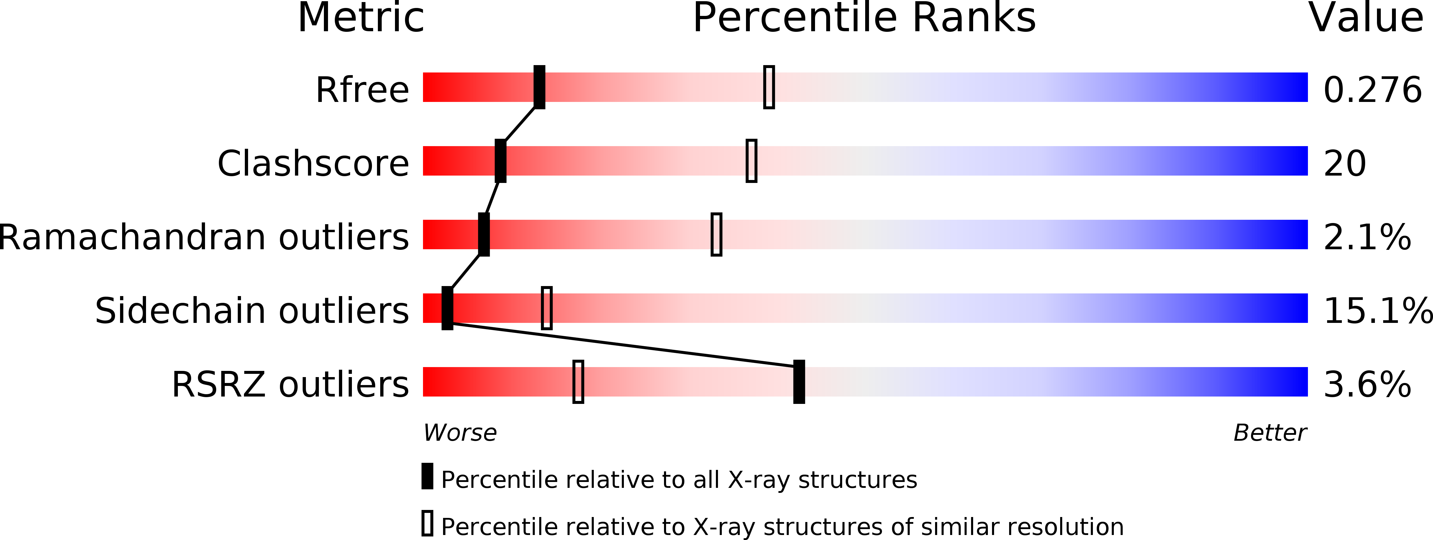
2WOO image
Deposition Date 2009-07-27
Release Date 2009-08-11
Last Version Date 2023-12-20
Entry Detail
PDB ID:
2WOO
Keywords:
Title:
Nucleotide-free form of S. pombe Get3
Biological Source:
Source Organism:
Host Organism:
Method Details:
Experimental Method:
Resolution:
3.01 Å
R-Value Free:
0.28
R-Value Work:
0.23
R-Value Observed:
0.23
Space Group:
P 21 21 21
Macromolecular Entities
Structures with similar UniProt ID
Protein Blast
Polymer Type:polypeptide(L)
Molecule:ATPASE GET3
Gene (Uniprot):get3
Chain IDs:A, B, C, D, E, F
Chain Length:329
Number of Molecules:6
Biological Source:SCHIZOSACCHAROMYCES POMBE
Ligand Molecules
Primary Citation
The Structural Basis of Tail-Anchored Membrane Protein Recognition by Get3.
Nature 461 361 ? (2009)
PMID: 19675567 DOI: 10.1038/NATURE08319

Abstact

Targeting of newly synthesized membrane proteins to the endoplasmic reticulum is an essential cellular process. Most membrane proteins are recognized and targeted co-translationally by the signal recognition particle. However, nearly 5% of membrane proteins are 'tail-anchored' by a single carboxy-terminal transmembrane domain that cannot access the co-translational pathway. Instead, tail-anchored proteins are targeted post-translationally by a conserved ATPase termed Get3. The mechanistic basis for tail-anchored protein recognition or targeting by Get3 is not known. Here we present crystal structures of yeast Get3 in 'open' (nucleotide-free) and 'closed' (ADP.AlF(4)(-)-bound) dimer states. In the closed state, the dimer interface of Get3 contains an enormous hydrophobic groove implicated by mutational analyses in tail-anchored protein binding. In the open state, Get3 undergoes a striking rearrangement that disrupts the groove and shields its hydrophobic surfaces. These data provide a molecular mechanism for nucleotide-regulated binding and release of tail-anchored proteins during their membrane targeting by Get3.

Legend

Protein

Chemical

Disease

Primary Citation of related structures