Deposition Date
2009-07-07
Release Date
2009-08-18
Last Version Date
2023-12-13
Entry Detail
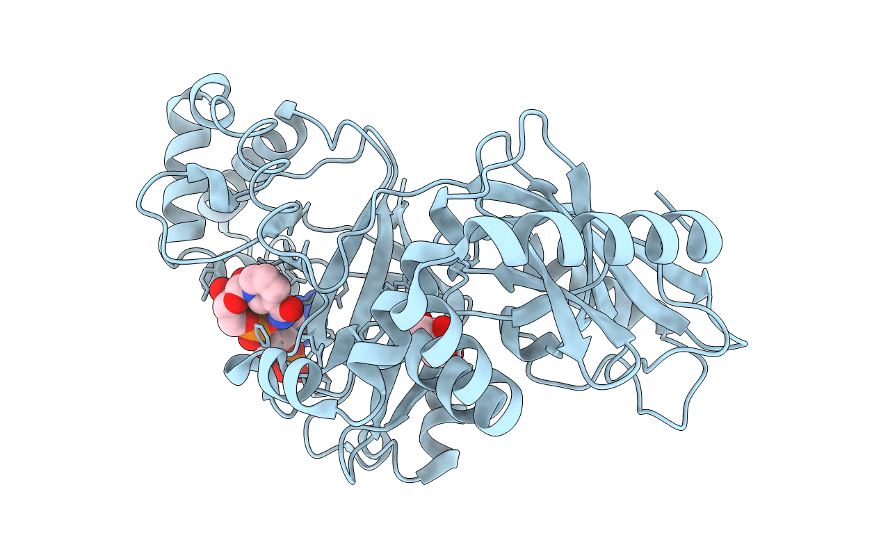
PDB ID:
2WN6
Keywords:
Title:
Structural Basis for Substrate Recognition in the Enzymatic Component of ADP-ribosyltransferase Toxin CDTa from Clostridium difficile
Biological Source:
Source Organism(s):
CLOSTRIDIUM DIFFICILE (Taxon ID: 1496)
Expression System(s):
Method Details:
Experimental Method:
Resolution:
1.96 Å
R-Value Free:
0.25
R-Value Work:
0.20
R-Value Observed:
0.20
Space Group:
P 1 21 1