2WN2 image
Deposition Date 2009-07-07
Release Date 2010-05-26
Last Version Date 2023-12-13
Entry Detail
PDB ID:
2WN2
Keywords:
Title:
Structure of the discoidin I from Dictyostelium discoideum in complex with galactose beta 1-3 galNAc at 1.8 A resolution.
Biological Source:
Source Organism:
Host Organism:
Method Details:
Experimental Method:
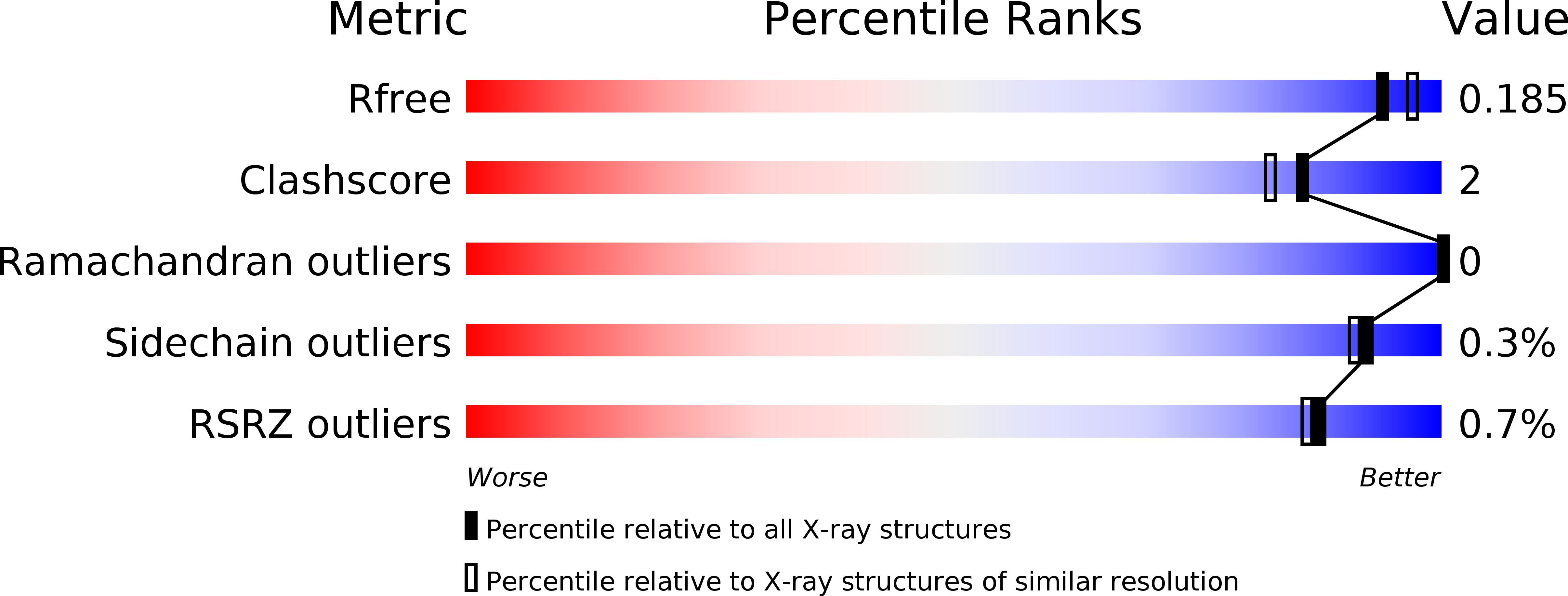
Resolution:
1.82 Å
R-Value Free:
0.18
R-Value Work:
0.14
R-Value Observed:
0.14
Space Group:
C 2 2 21
Macromolecular Entities
Polymer Type:polypeptide(L)
Molecule:DISCOIDIN-1 SUBUNIT A
Gene (Uniprot):dscA-1, dscA-2
Chain IDs:A, B, C
Chain Length:254
Number of Molecules:3
Biological Source:DICTYOSTELIUM DISCOIDEUM
Primary Citation
Discoidin I from Dictyostelium Discoideum and Interactions with Oligosaccharides: Specificity, Affinity, Crystal Structures and Comparison with Discoidin II.
J.Mol.Biol. 400 540 ? (2010)
PMID: 20580724 DOI: 10.1016/J.JMB.2010.05.042

Abstact

Discoidin I (DiscI) and discoidin II (DiscII) are N-acetylgalactosamine (GalNAc)-binding proteins from Dictyostelium discoideum. They consist of two domains: an N-terminal discoidin domain and a C-terminal H-type lectin domain. They were cloned and expressed in high yield in recombinant form in Escherichia coli. Although both lectins bind galactose (Gal) and GalNAc, glycan array experiments performed on the recombinant proteins displayed strong differences in their specificity for oligosaccharides. DiscI and DiscII bind preferentially to Gal/GalNAcbeta1-3Gal/GalNAc-containing and Gal/GalNAcbeta1-4GlcNAcbeta1-6Gal/GalNAc-containing glycans, respectively. The affinity of the interaction of DiscI with monosaccharides and disaccharides was evaluated using isothermal titration calorimetry experiments. The three-dimensional structures of native DiscI and its complexes with GalNAc, GalNAcbeta1-3Gal, and Galbeta1-3GalNAc were solved by X-ray crystallography. DiscI forms trimers with involvement of calcium at the monomer interface. The N-terminal discoidin domain presents a structural similarity to F-type lectins such as the eel agglutinin, where an amphiphilic binding pocket suggests possible carbohydrate-binding activity. In the C-terminal H-type lectin domain, the GalNAc residue establishes specific hydrogen bonds that explain the observed affinity (K(d)=3x10(-4) M). The different specificities of DiscI and DiscII for oligosaccharides were rationalized from the different structures obtained by either X-ray crystallography or molecular modeling.

Legend

Protein

Chemical

Disease

Primary Citation of related structures