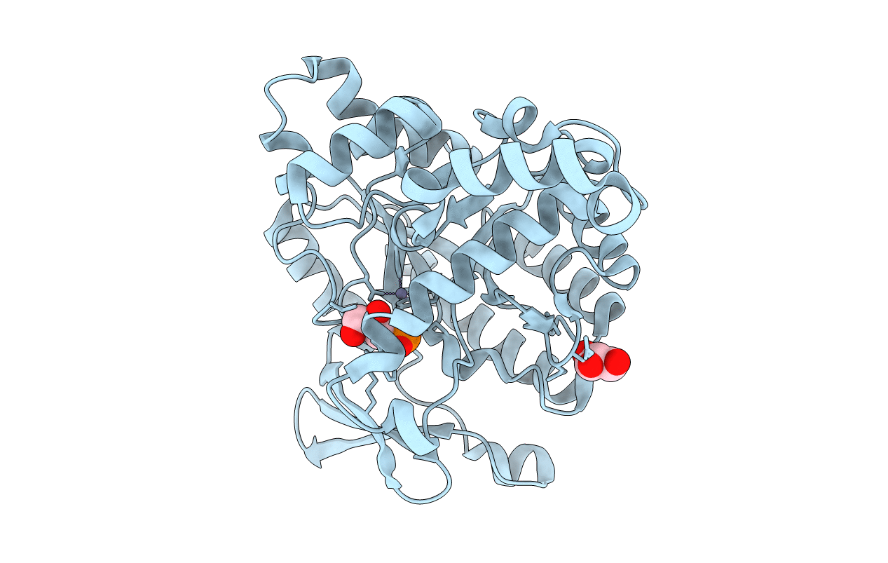
2WM1 image
Entry Detail
PDB ID:
2WM1
Keywords:
Title:
The crystal structure of human alpha-amino-beta-carboxymuconate- epsilon-semialdehyde decarboxylase in complex with 1,3- dihydroxyacetonephosphate suggests a regulatory link between NAD synthesis and glycolysis
Biological Source:
Source Organism:
Host Organism:
PDB Version:
Deposition Date:
2009-06-29
Release Date:
2009-11-03
Method Details:
Experimental Method:
Resolution:
2.01 Å
R-Value Free:
0.25
R-Value Work:
0.19
R-Value Observed:
0.19
Space Group:
P 32 2 1
Macromolecular Entities
Polymer Type:polypeptide(L)
Description:2-AMINO-3-CARBOXYMUCONATE-6-SEMIALDEHYDE DECARBOXYLASE
Chain IDs:A
Chain Length:336
Number of Molecules:1
Biological Source:HOMO SAPIENS
Primary Citation
The Crystal Structure of Human Alpha-Amino-Beta-Carboxymuconate-Epsilon-Semialdehyde Decarboxylase in Complex with 1,3-Dihydroxyacetonephosphate Suggests a Regulatory Link between Nad Synthesis and Glycolysis
FEBS J. 276 6615 ? (2009)
PMID: 19843166 DOI: 10.1111/J.1742-4658.2009.07372.X

Abstact

The enzyme alpha-amino-beta-carboxymuconate-epsilon-semialdehyde decarboxylase (ACMSD) is a zinc-dependent amidohydrolase that participates in picolinic acid (PA), quinolinic acid (QA) and NAD homeostasis. Indeed, the enzyme stands at a branch point of the tryptophan to NAD pathway, and determines the final fate of the amino acid, i.e. transformation into PA, complete oxidation through the citric acid cycle, or conversion into NAD through QA synthesis. Both PA and QA are key players in a number of physiological and pathological conditions, mainly affecting the central nervous system. As their relative concentrations must be tightly controlled, modulation of ACMSD activity appears to be a promising prospect for the treatment of neurological disorders, including cerebral malaria. Here we report the 2.0 A resolution crystal structure of human ACMSD in complex with the glycolytic intermediate 1,3-dihydroxyacetonephosphate (DHAP), refined to an R-factor of 0.19. DHAP, which we discovered to be a potent enzyme inhibitor, resides in the ligand binding pocket with its phosphate moiety contacting the catalytically essential zinc ion through mediation of a solvent molecule. Arg47, Asp291 and Trp191 appear to be the key residues for DHAP recognition in human ACMSD. Ligand binding induces a significant conformational change affecting a strictly conserved Trp-Met couple, and we propose that these residues are involved in controlling ligand admission into ACMSD. Our data may be used for the design of inhibitors with potential medical interest, and suggest a regulatory link between de novo NAD biosynthesis and glycolysis.

Legend

Protein

Chemical

Disease

Primary Citation of related structures