2WL7 image
Deposition Date 2009-06-22
Release Date 2010-01-19
Last Version Date 2023-12-13
Entry Detail
PDB ID:
2WL7
Keywords:
Title:
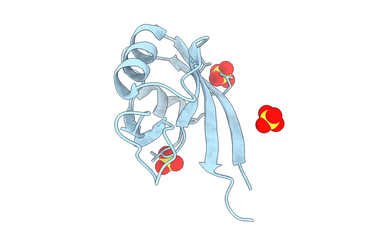
Crystal structure of the PSD93 PDZ1 domain
Biological Source:
Source Organism:
MUS MUSCULUS (Taxon ID: 10090)
Host Organism:
Method Details:
Experimental Method:
Resolution:
2.03 Å
R-Value Free:
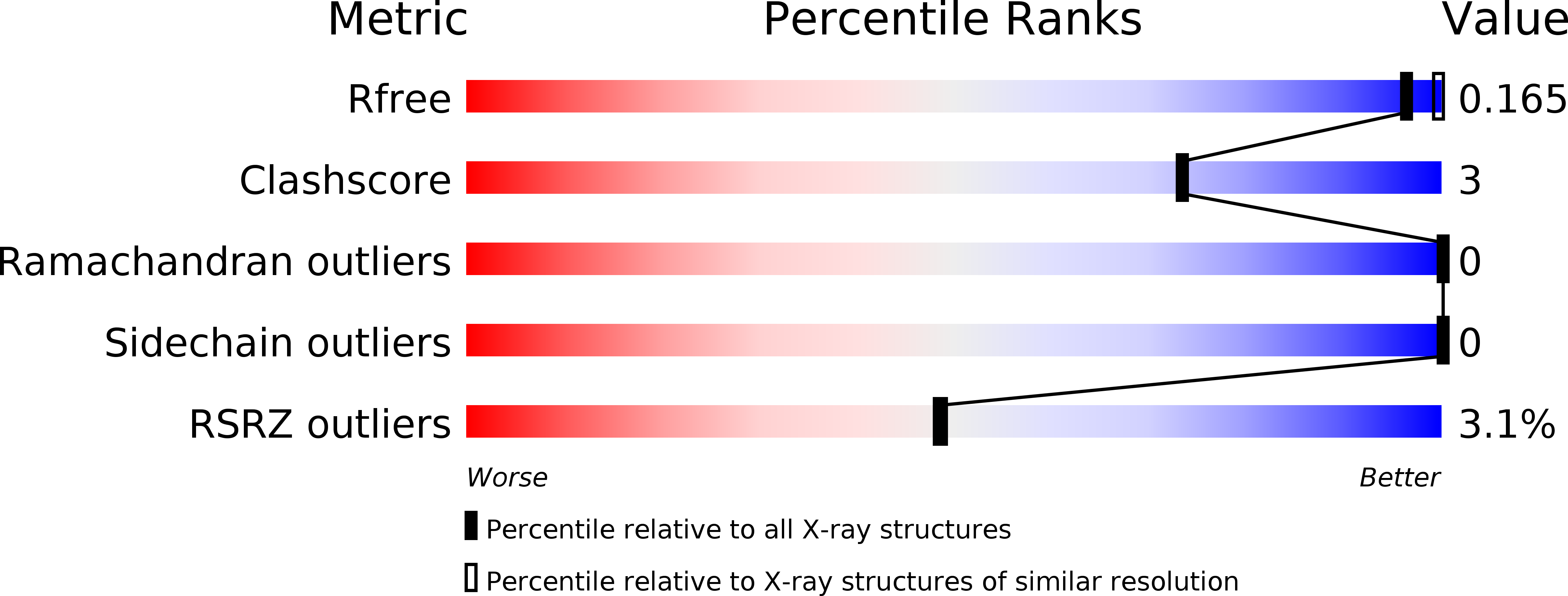
0.20
R-Value Work:
0.16
R-Value Observed:
0.16
Space Group:
H 3
Macromolecular Entities
Structures with similar UniProt ID
Protein Blast
Polymer Type:polypeptide(L)
Molecule:DISKS LARGE HOMOLOG 2
Gene (Uniprot):Dlg2
Chain IDs:A
Chain Length:102
Number of Molecules:1
Biological Source:MUS MUSCULUS
Primary Citation
Structure of the First Pdz Domain of Human Psd-93.
Acta Crystallogr.,Sect.F 65 1254 ? (2009)
PMID: 20054121 DOI: 10.1107/S1744309109043267

Abstact

The crystal structure of the PDZ1 domain of human PSD-93 has been determined to 2.0 A resolution. The PDZ1 domain forms a crystallographic trimer that is also predicted to be stable in solution. The main contributions to the stabilization of the trimer seem to arise from interactions involving the PDZ1-PDZ2 linker region at the extreme C-terminus of PDZ1, implying that the oligomerization that is observed is not of biological significance in full-length PSD-93. Comparison of the structures of the binding cleft of PSD-93 PDZ1 with the previously reported structures of PSD-93 PDZ2 and PDZ3 as well as of the closely related human PSD-95 PDZ1 shows that they are very similar in terms of amino-acid composition. However, the cleft is significantly narrower in PSD-95. This could be part of the basis of peptide selectivity between PSD-93 PDZ1 and PSD-95 PDZ1.

Legend

Protein

Chemical

Disease

Primary Citation of related structures