Deposition Date
2009-06-18
Release Date
2010-01-12
Last Version Date
2023-12-13
Entry Detail
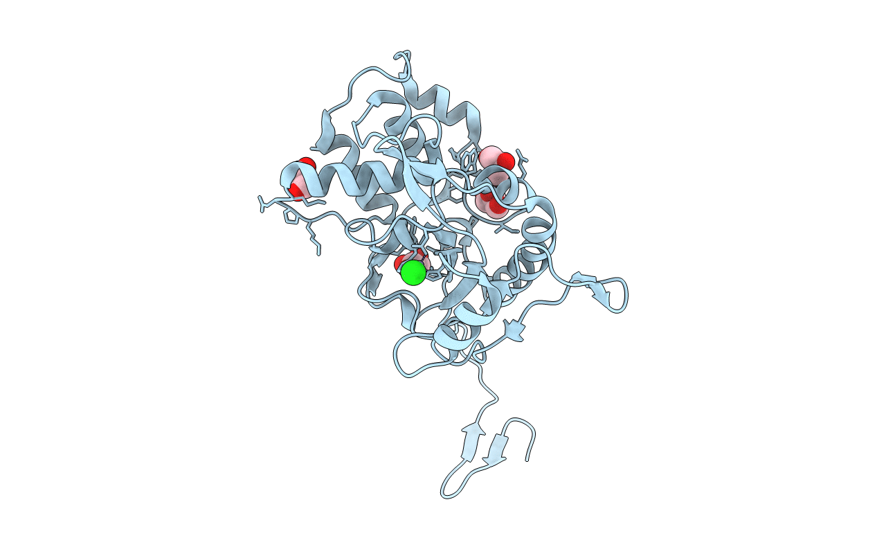
Biological Source:
Source Organism(s):
ESCHERICHIA COLI (Taxon ID: 83333)
Expression System(s):
Method Details:
Experimental Method:
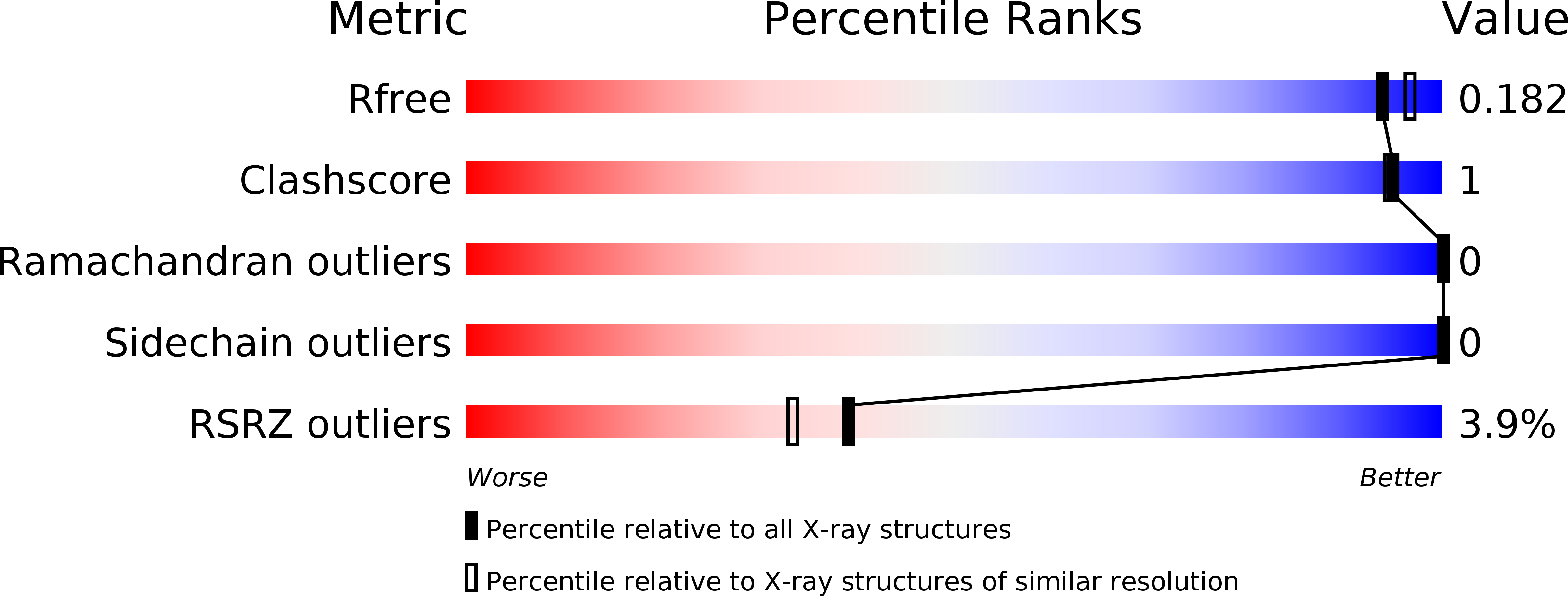
Resolution:
1.80 Å
R-Value Free:
0.18
R-Value Work:
0.16
R-Value Observed:
0.16
Space Group:
P 61 2 2