2WJS image
Deposition Date 2009-05-28
Release Date 2009-06-23
Last Version Date 2024-11-13
Entry Detail
PDB ID:
2WJS
Keywords:
Title:
Crystal structure of the LG1-3 region of the laminin alpha2 chain
Biological Source:
Source Organism:
MUS MUSCULUS (Taxon ID: 10090)
Host Organism:
Method Details:
Experimental Method:
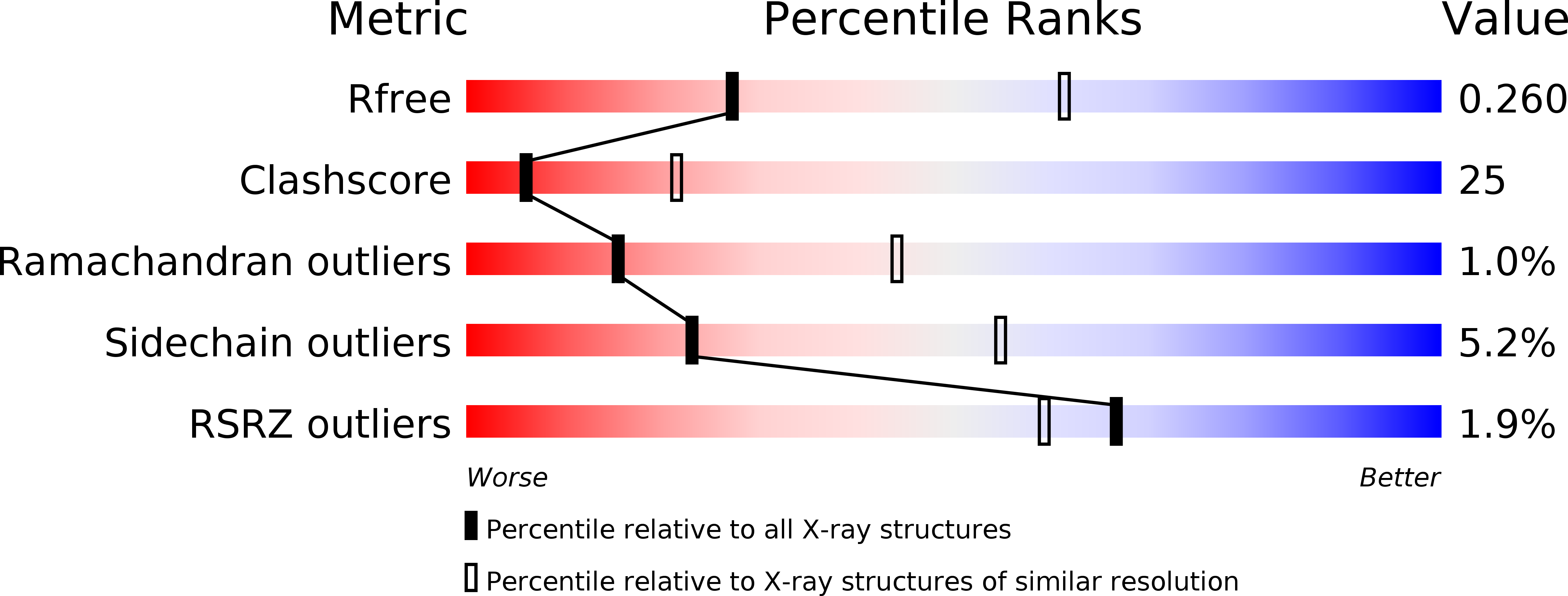
Resolution:
2.80 Å
R-Value Free:
0.26
R-Value Work:
0.21
R-Value Observed:
0.21
Space Group:
P 65
Macromolecular Entities
Structures with similar UniProt ID
Protein Blast
Polymer Type:polypeptide(L)
Molecule:LAMININ SUBUNIT ALPHA-2
Gene (Uniprot):Lama2
Chain IDs:A
Chain Length:608
Number of Molecules:1
Biological Source:MUS MUSCULUS
Modified Residue
Compound ID Chain ID Parent Comp ID Details 2D Image
ASN A ASN GLYCOSYLATION SITE
Primary Citation
Crystal Structure of the Lg1-3 Region of the Laminin {Alpha}2 Chain.
J.Biol.Chem. 284 22786 ? (2009)
PMID: 19553699 DOI: 10.1074/JBC.M109.026658

Abstact

Laminins are large heterotrimeric glycoproteins with many essential functions in basement membrane assembly and function. Cell adhesion to laminins is mediated by a tandem of five laminin G-like (LG) domains at the C terminus of the alpha chain. Integrin binding requires an intact LG1-3 region, as well as contributions from the coiled coil formed by the alpha, beta, and gamma chains. We have determined the crystal structure at 2.8-A resolution of the LG1-3 region of the laminin alpha2 chain (alpha 2LG1-3). The three LG domains adopt typical beta-sandwich folds, with canonical calcium binding sites in LG1 and LG2. LG2 and LG3 interact through a substantial interface, but LG1 is completely dissociated from the LG2-3 pair. We suggest that the missing gamma chain tail may be required to stabilize the interaction between LG1 and LG2-3 in the biologically active conformation. A global analysis of N-linked glycosylation sites shows that the beta-sandwich faces of LG1 are free of carbohydrate modifications in all five laminin alpha chains, suggesting that these surfaces may harbor the integrin binding site. The alpha 2LG1-3 structure provides the first atomic view of the integrin binding region of laminins.

Legend

Protein

Chemical

Disease

Primary Citation of related structures