2WHV image
Deposition Date 2009-05-07
Release Date 2010-04-21
Last Version Date 2023-12-13
Entry Detail
PDB ID:
2WHV
Keywords:
Title:
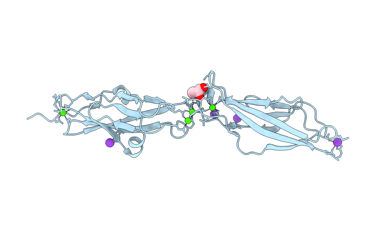
CRYSTAL STRUCTURE OF MOUSE CADHERIN-23 EC1-2 (ALL CATION BINDING SITES OCCUPIED BY CALCIUM)
Biological Source:
Source Organism:
MUS MUSCULUS (Taxon ID: 10090)
Host Organism:
Method Details:
Experimental Method:
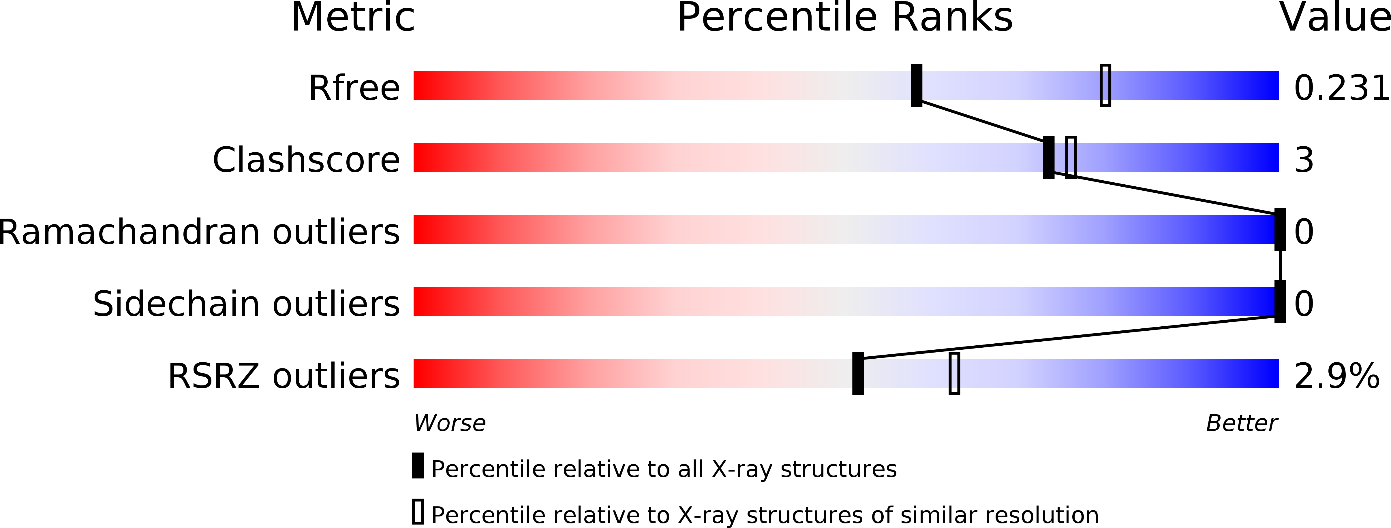
Resolution:
2.36 Å
R-Value Free:
0.22
R-Value Work:
0.19
R-Value Observed:
0.20
Space Group:
H 3 2
Macromolecular Entities
Structures with similar UniProt ID
Protein Blast
Polymer Type:polypeptide(L)
Molecule:CADHERIN-23
Gene (Uniprot):Cdh23
Chain IDs:A
Chain Length:214
Number of Molecules:1
Biological Source:MUS MUSCULUS
Primary Citation
Structural Determinants of Cadherin-23 Function in Hearing and Deafness.
Neuron 66 85 ? (2010)
PMID: 20399731 DOI: 10.1016/J.NEURON.2010.03.028

Abstact

The hair-cell tip link, a fine filament directly conveying force to mechanosensitive transduction channels, is composed of two proteins, protocadherin-15 and cadherin-23, whose mutation causes deafness. However, their molecular structure, elasticity, and deafness-related structural defects are unknown. We present crystal structures of the first and second extracellular cadherin repeats of cadherin-23. Overall, structures show typical cadherin folds, but reveal an elongated N terminus that precludes classical cadherin interactions and contributes to an N-terminal Ca(2+)-binding site. The deafness mutation D101G, in the linker region between the repeats, causes a slight bend between repeats and decreases Ca(2+) affinity. Molecular dynamics simulations suggest that cadherin-23 repeats are stiff and that either removing Ca(2+) or mutating Ca(2+)-binding residues reduces rigidity and unfolding strength. The structures define an uncharacterized cadherin family and, with simulations, suggest mechanisms underlying inherited deafness and how cadherin-23 may bind with itself and with protocadherin-15 to form the tip link.

Legend

Protein

Chemical

Disease

Primary Citation of related structures