2WHP image
Entry Detail
PDB ID:
2WHP
Keywords:
Title:
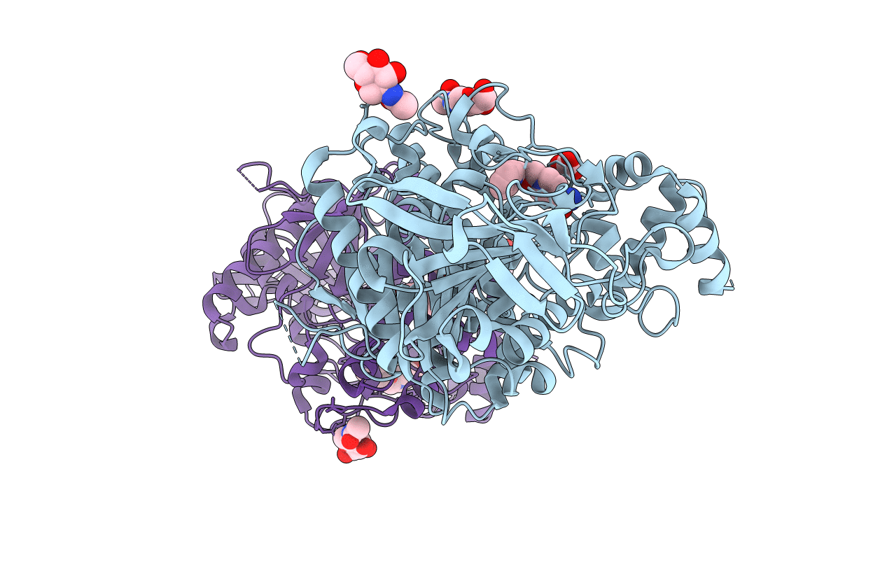
Crystal structure of acetylcholinesterase, phosphonylated by sarin and in complex with HI-6
Biological Source:
Source Organism:
Host Organism:
PDB Version:
Deposition Date:
2009-05-06
Release Date:
2009-06-30
Method Details:
Experimental Method:
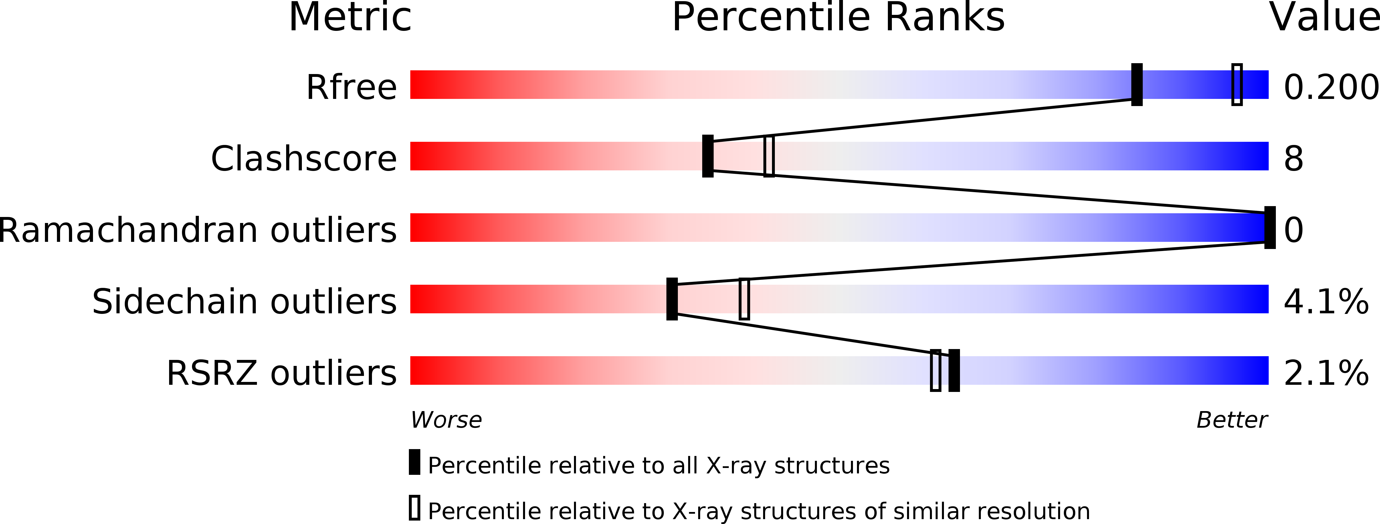
Resolution:
2.20 Å
R-Value Free:
0.21
R-Value Work:
0.17
R-Value Observed:
0.17
Space Group:
P 21 21 21
Macromolecular Entities
Polymer Type:polypeptide(L)
Description:ACETYLCHOLINESTERASE
Chain IDs:A
Chain Length:548
Number of Molecules:1
Biological Source:MUS MUSCULUS
Polymer Type:polypeptide(L)
Description:ACETYLCHOLINESTERASE
Chain IDs:B
Chain Length:548
Number of Molecules:1
Biological Source:MUS MUSCULUS
Modified Residue
Compound ID Chain ID Parent Comp ID Details 2D Image
ASN A ASN GLYCOSYLATION SITE
SGB A SER ?
Primary Citation
Structure of Hi-6Sarin-Acetylcholinesterase Determined by X-Ray Crystallography and Molecular Dynamics Simulation: Reactivator Mechanism and Design.
Plos One 4 E5957 ? (2009)
PMID: 19536291 DOI: 10.1371/JOURNAL.PONE.0005957

Abstact

Organophosphonates such as isopropyl metylphosphonofluoridate (sarin) are extremely toxic as they phosphonylate the catalytic serine residue of acetylcholinesterase (AChE), an enzyme essential to humans and other species. Design of effective AChE reactivators as antidotes to various organophosphonates requires information on how the reactivators interact with the phosphonylated AChEs. However, such information has not been available hitherto because of three main challenges. First, reactivators are generally flexible in order to change from the ground state to the transition state for reactivation; this flexibility discourages determination of crystal structures of AChE in complex with effective reactivators that are intrinsically disordered. Second, reactivation occurs upon binding of a reactivator to the phosphonylated AChE. Third, the phosphorous conjugate can develop resistance to reactivation. We have identified crystallographic conditions that led to the determination of a crystal structure of the sarin(nonaged)-conjugated mouse AChE in complex with [(E)-[1-[(4-carbamoylpyridin-1-ium-1-yl)methoxymethyl]pyridin-2-ylidene]methyl]-oxoazanium dichloride (HI-6) at a resolution of 2.2 A. In this structure, the carboxyamino-pyridinium ring of HI-6 is sandwiched by Tyr124 and Trp286, however, the oxime-pyridinium ring is disordered. By combining crystallography with microsecond molecular dynamics simulation, we determined the oxime-pyridinium ring structure, which shows that the oxime group of HI-6 can form a hydrogen-bond network to the sarin isopropyl ether oxygen, and a water molecule is able to form a hydrogen bond to the catalytic histidine residue and subsequently deprotonates the oxime for reactivation. These results offer insights into the reactivation mechanism of HI-6 and design of better reactivators.

Legend

Protein

Chemical

Disease