2WGF image
Entry Detail
PDB ID:
2WGF
Keywords:
Title:
Crystal structure of Mycobacterium tuberculosis C171Q KasA variant
Biological Source:
PDB Version:
Deposition Date:
2009-04-17
Release Date:
2009-07-21
Method Details:
Experimental Method:
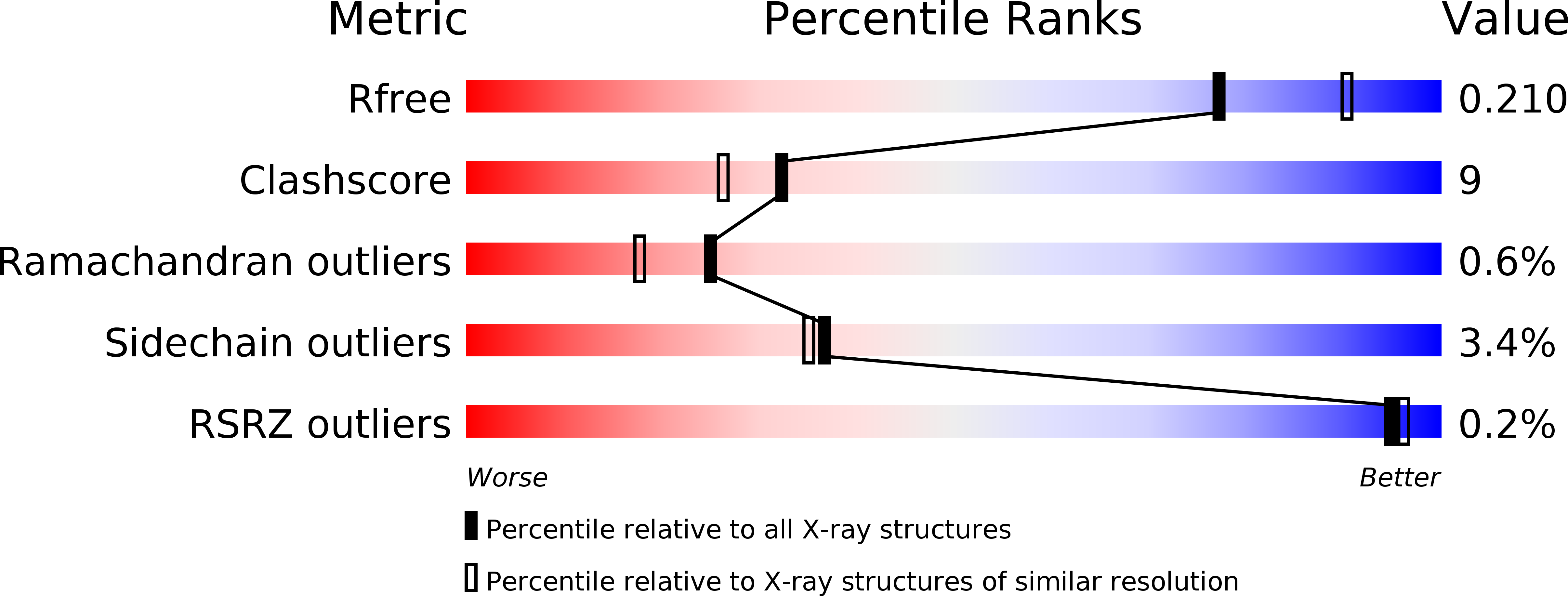
Resolution:
2.15 Å
R-Value Free:
0.21
R-Value Work:
0.16
R-Value Observed:
0.16
Space Group:
P 31
Macromolecular Entities
Polymer Type:polypeptide(L)
Description:3-OXOACYL-[ACYL-CARRIER-PROTEIN] SYNTHASE 1
Mutations:YES
Chain IDs:A, B, C, D, E, F, G, H
Chain Length:416
Number of Molecules:8
Biological Source:MYCOBACTERIUM TUBERCULOSIS
Primary Citation
Crystal Structures of Mycobacterium Tuberculosis Kasa Show Mode of Action within Cell Wall Biosynthesis and its Inhibition by Thiolactomycin
Structure 17 1004 ? (2009)
PMID: 19604480 DOI: 10.1016/J.STR.2009.04.012

Abstact

Mycobacteria have a unique cell wall consisting of mycolic acids, very-long-chain lipids that provide protection and allow the bacteria to persist within human macrophages. Inhibition of cell wall biosynthesis is fatal for the organism and a starting point for the discovery and development of novel antibiotics. We determined the crystal structures of KasA, a key enzyme involved in the biosynthesis of long-chain fatty acids, in its apo-form and bound to the natural product inhibitor thiolactomycin. Detailed insights into the interaction of the inhibitor with KasA and the identification of a polyethylene glycol molecule that mimics a fatty acid substrate of approximately 40 carbon atoms length, represent the first atomic view of a mycobacterial enzyme involved in the synthesis of long-chain fatty acids and provide a robust platform for the development of novel thiolactomycin analogs with high affinity for KasA.

Legend

Protein

Chemical

Disease

Primary Citation of related structures