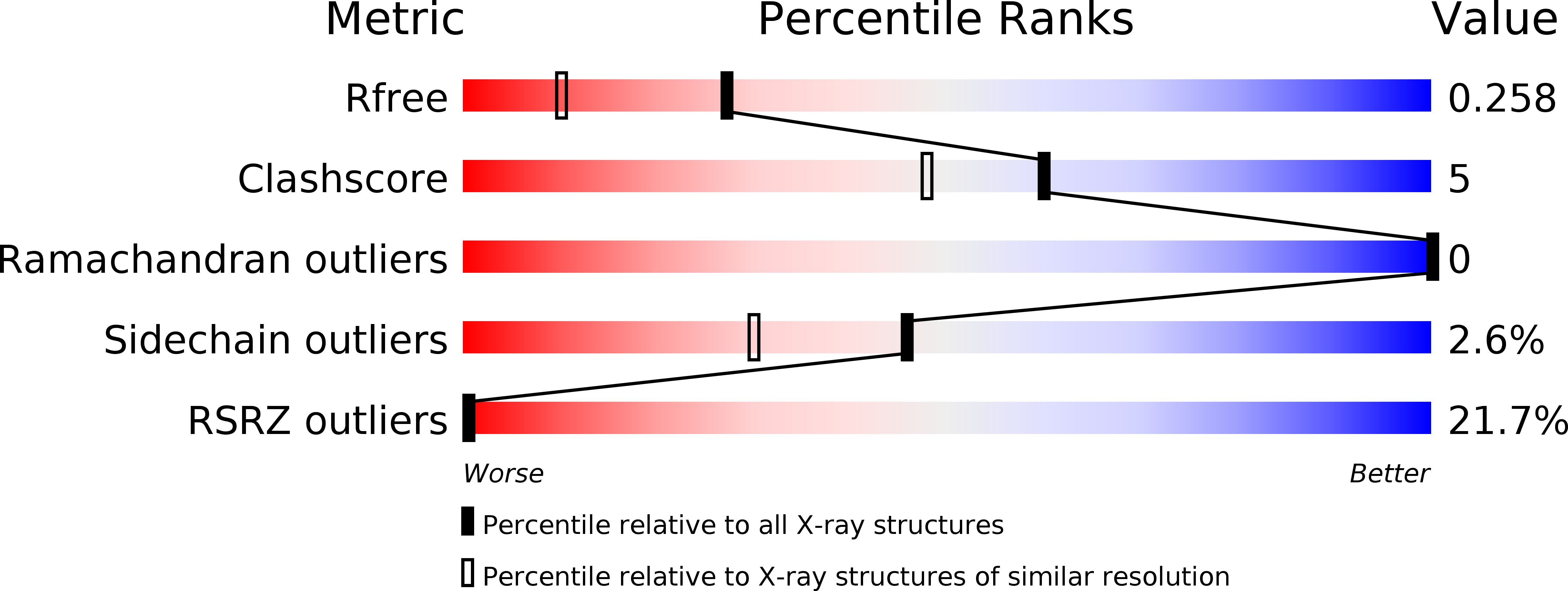
2WFU image
Deposition Date 2009-04-15
Release Date 2010-05-26
Last Version Date 2024-10-23
Entry Detail
PDB ID:
2WFU
Title:
Crystal structure of DILP5 variant DB
Biological Source:
Source Organism:
Method Details:
Experimental Method:
Resolution:
1.85 Å
R-Value Free:
0.25
R-Value Work:
0.21
R-Value Observed:
0.21
Space Group:
P 43 21 2
Macromolecular Entities
Polymer Type:polypeptide(L)
Molecule:PROBABLE INSULIN-LIKE PEPTIDE 5 A CHAIN
Gene (Uniprot):Ilp5
Mutations:YES
Chain IDs:A
Chain Length:22
Number of Molecules:1
Biological Source:DROSOPHILA MELANOGASTER
Polymer Type:polypeptide(L)
Molecule:PROBABLE INSULIN-LIKE PEPTIDE 5 B CHAIN
Gene (Uniprot):Ilp5
Chain IDs:B
Chain Length:24
Number of Molecules:1
Biological Source:DROSOPHILA MELANOGASTER
Primary Citation
Structural and Biological Properties of the Drosophila Insulin-Like Peptide 5 Show Evolutionary Conservation.
J.Biol.Chem. 286 661 ? (2011)
PMID: 20974844 DOI: 10.1074/JBC.M110.156018

Abstact

We report the crystal structure of two variants of Drosophila melanogaster insulin-like peptide 5 (DILP5) at a resolution of 1.85 Å. DILP5 shares the basic fold of the insulin peptide family (T conformation) but with a disordered B-chain C terminus. DILP5 dimerizes in the crystal and in solution. The dimer interface is not similar to that observed in vertebrates, i.e. through an anti-parallel β-sheet involving the B-chain C termini but, in contrast, is formed through an anti-parallel β-sheet involving the B-chain N termini. DILP5 binds to and activates the human insulin receptor and lowers blood glucose in rats. It also lowers trehalose levels in Drosophila. Reciprocally, human insulin binds to the Drosophila insulin receptor and induces negative cooperativity as in the human receptor. DILP5 also binds to insect insulin-binding proteins. These results show high evolutionary conservation of the insulin receptor binding properties despite divergent insulin dimerization mechanisms.

Legend

Protein

Chemical

Disease

Primary Citation of related structures