Deposition Date
2009-04-06
Release Date
2009-06-16
Last Version Date
2025-04-09
Entry Detail
PDB ID:
2WFJ
Keywords:
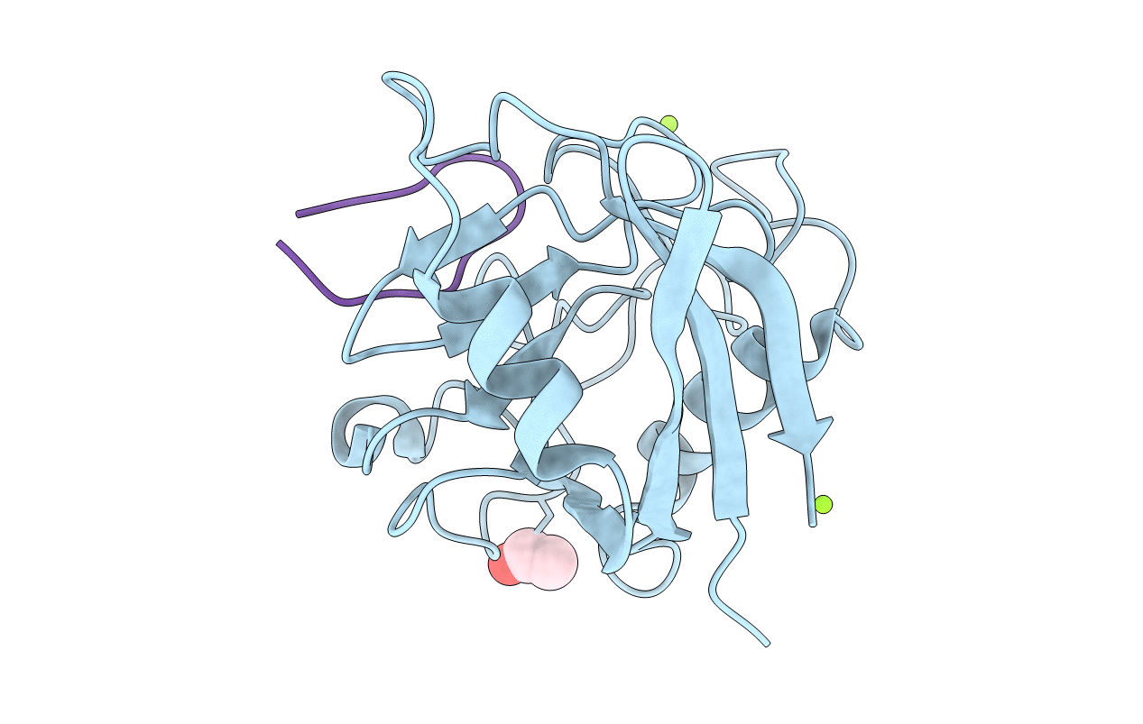
Title:
Atomic resolution crystal structure of the PPIase domain of human cyclophilin G in complex with cyclosporin A.
Biological Source:
Source Organism(s):
HOMO SAPIENS (Taxon ID: 9606)
TOLYPOCLADIUM INFLATUM (Taxon ID: 29910)
TOLYPOCLADIUM INFLATUM (Taxon ID: 29910)
Expression System(s):
Method Details:
Experimental Method:
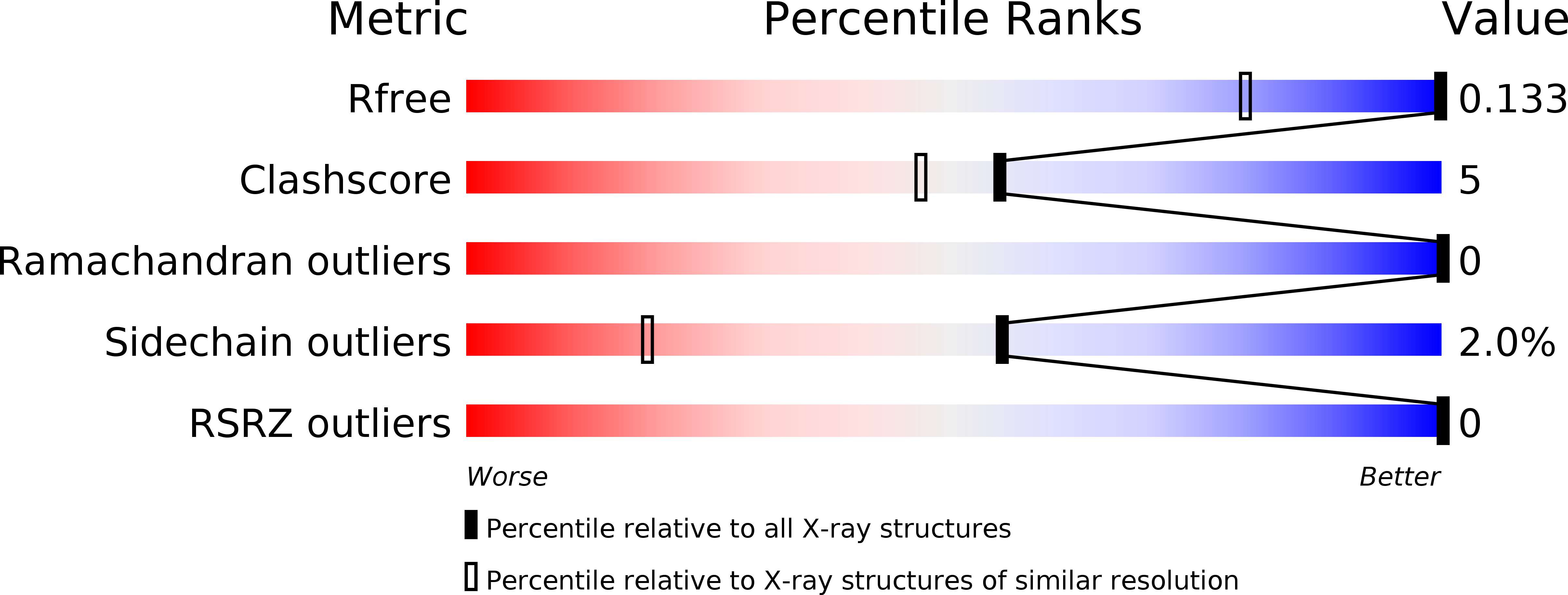
Resolution:
0.75 Å
R-Value Free:
0.12
R-Value Observed:
0.11
Space Group:
P 21 21 21