2WDR image
Entry Detail
PDB ID:
2WDR
Keywords:
Title:
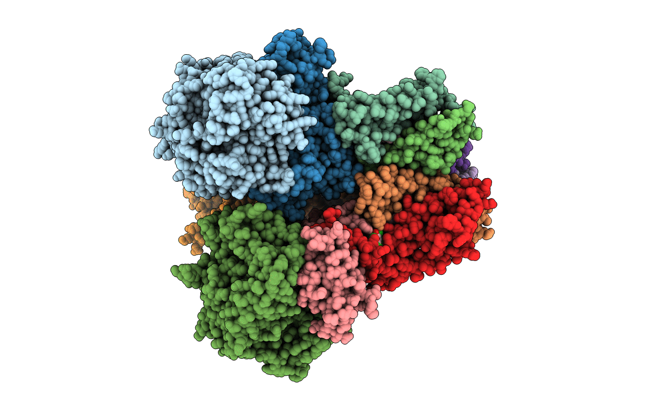
E. coli succinate:quinone oxidoreductase (SQR) with pentachlorophenol bound
Biological Source:
Source Organism:
Host Organism:
PDB Version:
Deposition Date:
2009-03-25
Release Date:
2009-08-25
Method Details:
Experimental Method:
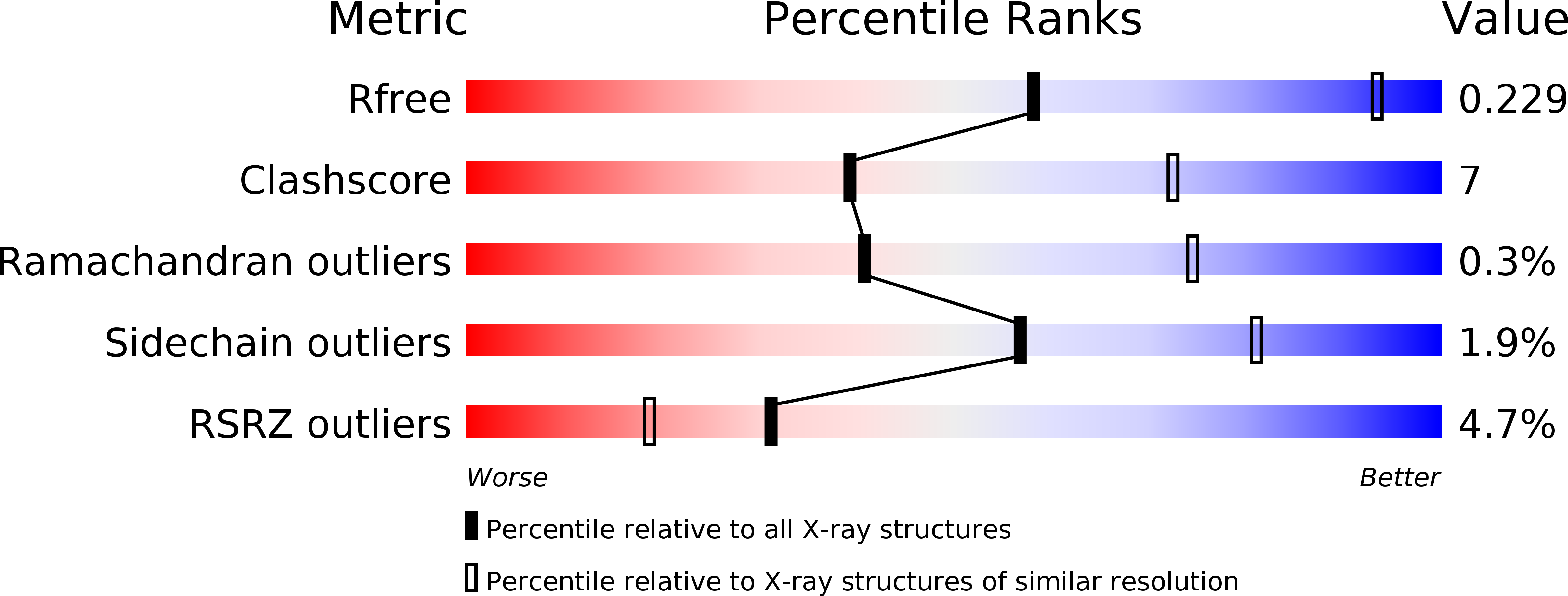
Resolution:
3.20 Å
R-Value Free:
0.22
R-Value Work:
0.19
R-Value Observed:
0.19
Space Group:
P 21 21 21
Macromolecular Entities
Polymer Type:polypeptide(L)
Description:SUCCINATE DEHYDROGENASE FLAVOPROTEIN SUBUNIT
Chain IDs:A, E, I
Chain Length:588
Number of Molecules:3
Biological Source:ESCHERICHIA COLI
Polymer Type:polypeptide(L)
Description:SUCCINATE DEHYDROGENASE IRON-SULFUR SUBUNIT
Chain IDs:B, F, J
Chain Length:238
Number of Molecules:3
Biological Source:ESCHERICHIA COLI
Polymer Type:polypeptide(L)
Description:SUCCINATE DEHYDROGENASE CYTOCHROME B556 SUBUNIT
Chain IDs:C, G, K
Chain Length:129
Number of Molecules:3
Biological Source:ESCHERICHIA COLI
Polymer Type:polypeptide(L)
Description:SUCCINATE DEHYDROGENASE HYDROPHOBIC MEMBRANE ANCHOR SUBUNIT
Chain IDs:D, H, L
Chain Length:115
Number of Molecules:3
Biological Source:ESCHERICHIA COLI
Primary Citation
Structure of Escherichia Coli Succinate:Quinone Oxidoreductase with an Occupied and Empty Quinone- Binding Site.
J.Biol.Chem. 284 29836 ? (2009)
PMID: 19710024 DOI: 10.1074/JBC.M109.010058

Abstact

Three new structures of Escherichia coli succinate-quinone oxidoreductase (SQR) have been solved. One with the specific quinone-binding site (Q-site) inhibitor carboxin present has been solved at 2.4 A resolution and reveals how carboxin inhibits the Q-site. The other new structures are with the Q-site inhibitor pentachlorophenol and with an empty Q-site. These structures reveal important details unresolved in earlier structures. Comparison of the new SQR structures shows how subtle rearrangements of the quinone-binding site accommodate the different inhibitors. The position of conserved water molecules near the quinone binding pocket leads to a reassessment of possible water-mediated proton uptake networks that complete reduction of ubiquinone. The dicarboxylate-binding site in the soluble domain of SQR is highly similar to that seen in high resolution structures of avian SQR (PDB 2H88) and soluble flavocytochrome c (PDB 1QJD) showing mechanistically significant structural features conserved across prokaryotic and eukaryotic SQRs.

Legend

Protein

Chemical

Disease

Primary Citation of related structures