2WD6 image
Deposition Date 2009-03-20
Release Date 2009-07-28
Last Version Date 2024-05-08
Entry Detail
PDB ID:
2WD6
Keywords:
Title:
Crystal structure of the Variable Domain of the Streptococcus gordonii Surface Protein SspB
Biological Source:
Source Organism:
Host Organism:
Method Details:
Experimental Method:
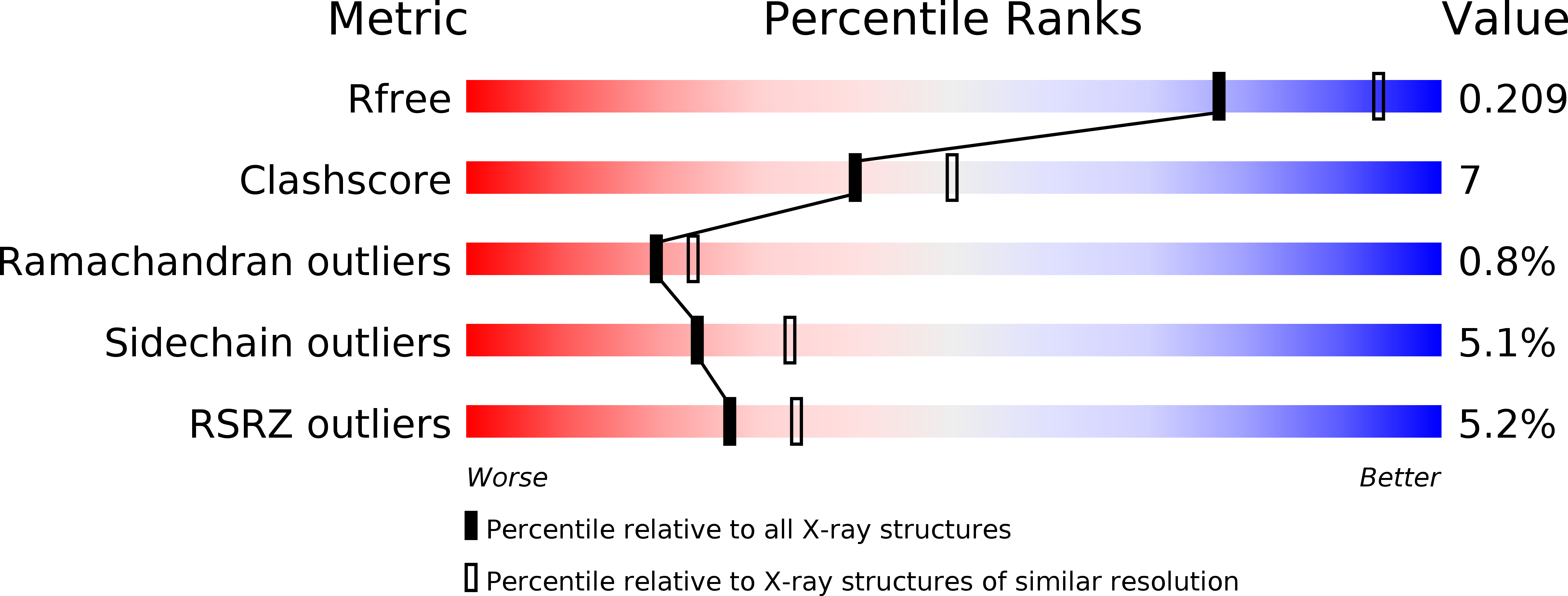
Resolution:
2.30 Å
R-Value Free:
0.20
R-Value Work:
0.17
R-Value Observed:
0.17
Space Group:
P 65
Macromolecular Entities
Polymer Type:polypeptide(L)
Molecule:AGGLUTININ RECEPTOR
Gene (Uniprot):sspB
Chain IDs:A, B
Chain Length:349
Number of Molecules:2
Biological Source:STREPTOCOCCUS GORDONII
Primary Citation
Crystal Structure of the Variable Domain of the Streptococcus Gordonii Surface Protein Sspb.
Protein Sci. 18 1896 ? (2009)
PMID: 19609934 DOI: 10.1002/PRO.200

Abstact

The Antigen I/II (AgI/II) family of proteins are cell wall anchored adhesins expressed on the surface of oral streptococci. The AgI/II proteins interact with molecules on other bacteria, on the surface of host cells, and with salivary proteins. Streptococcus gordonii is a commensal bacterium, and one of the primary colonizers that initiate the formation of the oral biofilm. S. gordonii expresses two AgI/II proteins, SspA and SspB that are closely related. One of the domains of SspB, called the variable (V-) domain, is significantly different from corresponding domains in SspA and all other AgI/II proteins. As a first step to elucidate the differences among these proteins, we have determined the crystal structure of the V-domain from S. gordonii SspB at 2.3 A resolution. The domain comprises a beta-supersandwich with a putative binding cleft stabilized by a metal ion. The overall structure of the SspB V-domain is similar to the previously reported V-domain of the Streptococcus mutans protein SpaP, despite their low sequence similarity. In spite of the conserved architecture of the binding cleft, the cavity is significantly smaller in SspB, which may provide clues about the difference in ligand specificity. We also verified that the metal in the binding cleft is a calcium ion, in concurrence with previous biological data. It was previously suggested that AgI/II V-domains are carbohydrate binding. However, we tested that hypothesis by screening the SspB V-domain for binding to over 400 glycoconjucates and found that the domain does not interact with any of the carbohydrates.

Legend

Protein

Chemical

Disease

Primary Citation of related structures