2W9Y image
Deposition Date 2009-01-30
Release Date 2009-10-20
Last Version Date 2025-04-09
Entry Detail
PDB ID:
2W9Y
Keywords:
Title:
The structure of the lipid binding protein Ce-FAR-7 from Caenorhabditis elegans
Biological Source:
Source Organism:
Method Details:
Experimental Method:
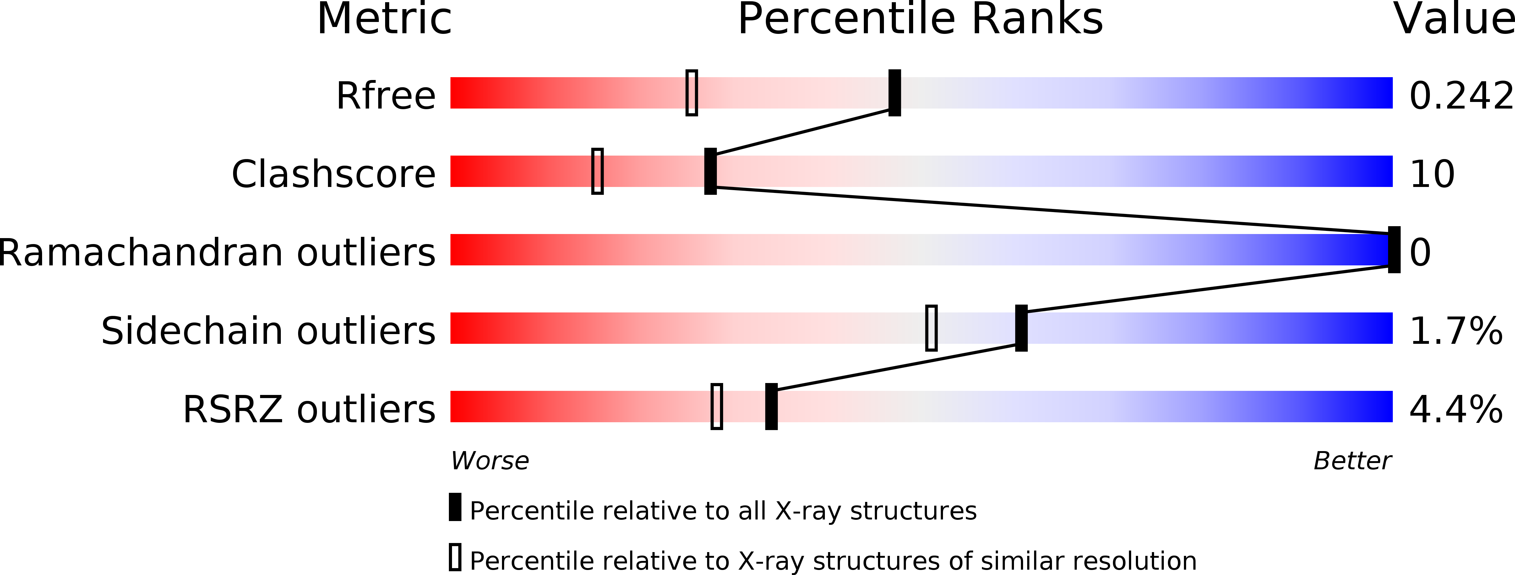
Resolution:
1.80 Å
R-Value Free:
0.25
R-Value Work:
0.17
R-Value Observed:
0.17
Space Group:
P 21 21 21
Macromolecular Entities
Structures with similar UniProt ID
Protein Blast
Polymer Type:polypeptide(L)
Molecule:FATTY ACID/RETINOL BINDING PROTEIN PROTEIN 7, ISOFORM A, CONFIRMED BY TRANSCRIPT EVIDENCE
Gene (Uniprot):far-7
Chain IDs:A
Chain Length:140
Number of Molecules:1
Biological Source:CAENORHABDITIS ELEGANS
Modified Residue
Compound ID Chain ID Parent Comp ID Details 2D Image
CSX A CYS S-OXY CYSTEINE
Ligand Molecules
Primary Citation
Fatty Acid and Retinoid Binding Proteins Have Distinct Binding Pockets for the Two Types of Cargo
J.Biol.Chem. 284 35818 ? (2009)
PMID: 19828452 DOI: 10.1074/JBC.M109.022731

Abstact

Parasitic nematodes cause serious diseases in humans, animals, and plants. They have limited lipid metabolism and are reliant on lipid-binding proteins to acquire these metabolites from their hosts. Several structurally novel families of lipid-binding proteins in nematodes have been described, including the fatty acid- and retinoid-binding protein family (FAR). In Caenorhabditis elegans, used as a model for studying parasitic nematodes, eight C. elegans FAR proteins have been described. The crystal structure of C. elegans FAR-7 is the first structure of a FAR protein, and it exhibits a novel fold. It differs radically from the mammalian fatty acid-binding proteins and has two ligand binding pockets joined by a surface groove. The first can accommodate the aliphatic chain of fatty acids, whereas the second can accommodate the bulkier retinoids. In addition to demonstrating lipid binding by fluorescence spectroscopy, we present evidence that retinol binding is positively regulated by casein kinase II phosphorylation at a conserved site near the bottom of the second pocket. far-7::GFP (green fluorescent protein) expression shows that it is localized in the head hypodermal syncytia and the excretory cell but that this localization changes under starvation conditions. In conclusion, our study provides the basic structural and functional information for investigation of inhibitors of lipid binding by FAR proteins.

Legend

Protein

Chemical

Disease

Primary Citation of related structures