Deposition Date
2009-01-25
Release Date
2009-02-03
Last Version Date
2024-10-16
Entry Detail
PDB ID:
2W9J
Keywords:
Title:
The crystal structure of SRP14 from the Schizosaccharomyces pombe signal recognition particle
Biological Source:
Source Organism(s):
SCHIZOSACCHAROMYCES POMBE (Taxon ID: 4896)
Expression System(s):
Method Details:
Experimental Method:
Resolution:
2.60 Å
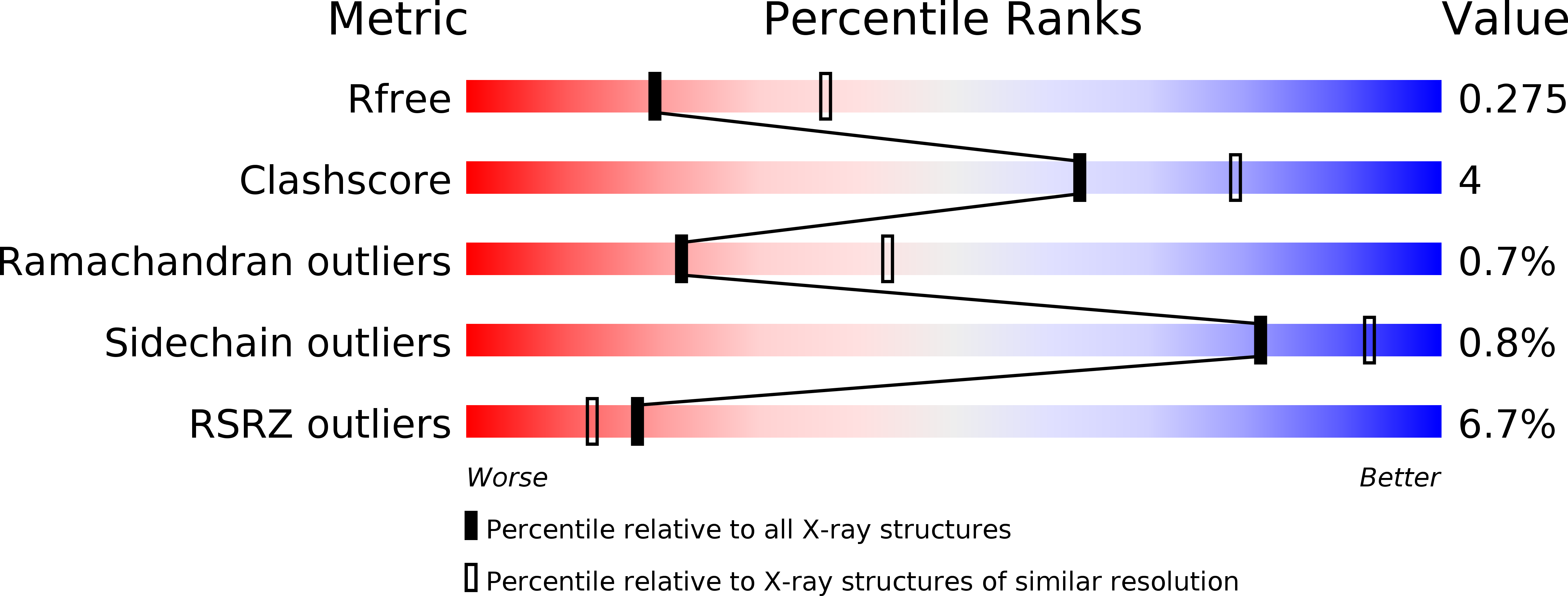
R-Value Free:
0.28
R-Value Work:
0.23
R-Value Observed:
0.24
Space Group:
P 32 2 1