Deposition Date
2009-01-22
Release Date
2010-03-31
Last Version Date
2023-12-13
Entry Detail
PDB ID:
2W97
Keywords:
Title:
Crystal Structure of eIF4E Bound to Glycerol and eIF4G1 peptide
Biological Source:
Source Organism(s):
HOMO SAPIENS (Taxon ID: 9606)
Expression System(s):
Method Details:
Experimental Method:
Resolution:
2.29 Å
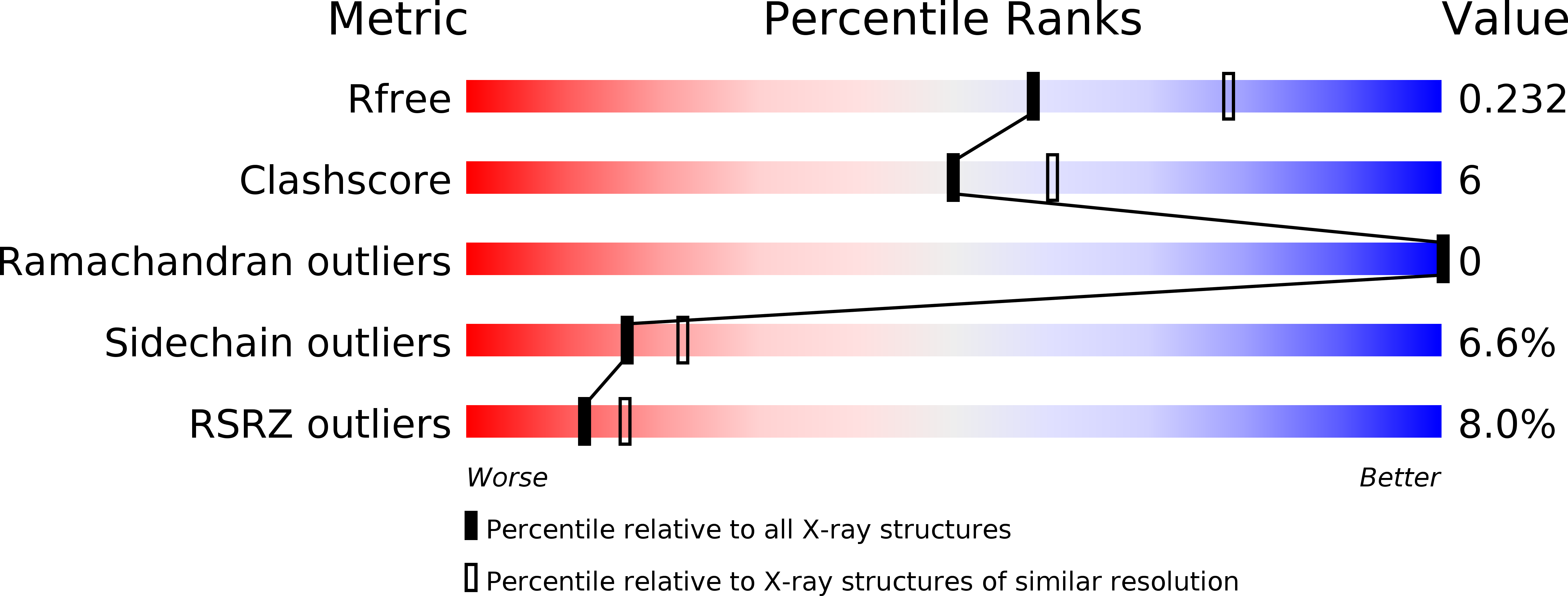
R-Value Free:
0.23
R-Value Work:
0.19
R-Value Observed:
0.19
Space Group:
P 1 21 1