Deposition Date
2008-12-19
Release Date
2009-04-28
Last Version Date
2024-02-07
Entry Detail
PDB ID:
2W72
Keywords:
Title:
DEOXYGENATED STRUCTURE OF A DISTAL SITE HEMOGLOBIN MUTANT PLUS XE
Biological Source:
Source Organism(s):
HOMO SAPIENS (Taxon ID: 9606)
Expression System(s):
Method Details:
Experimental Method:
Resolution:
1.07 Å
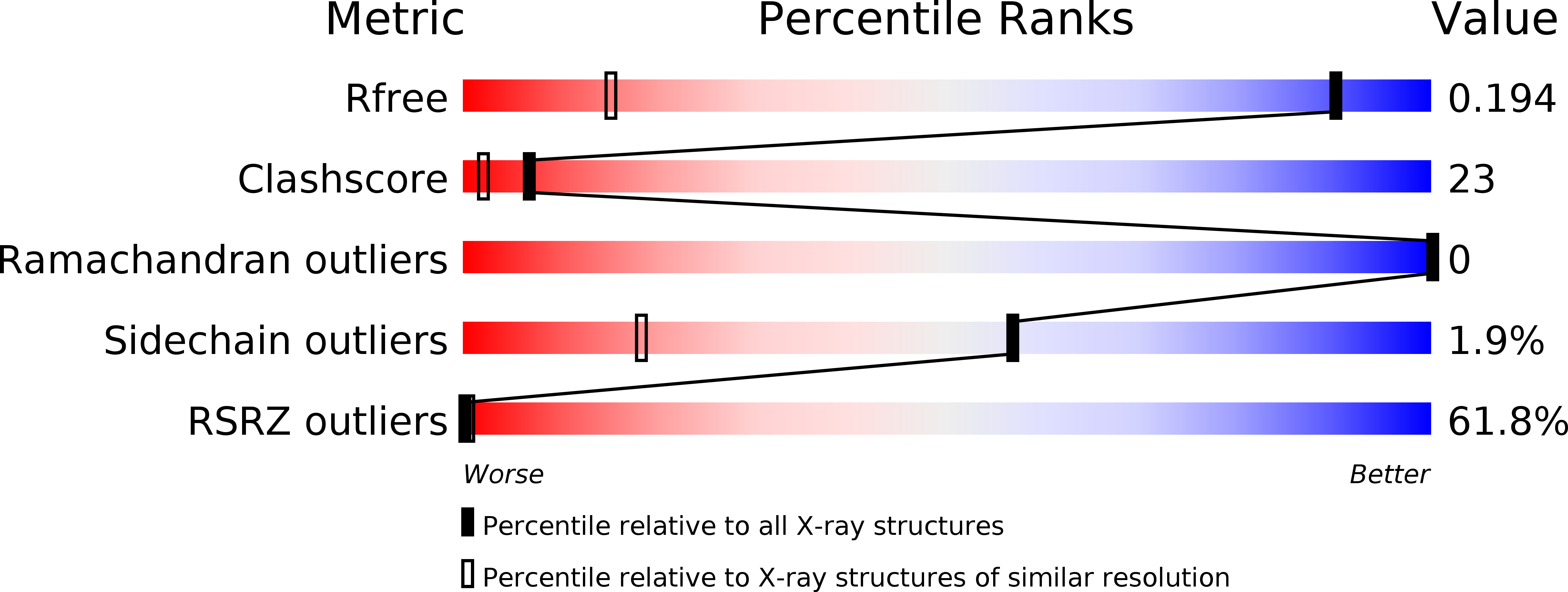
R-Value Free:
0.15
R-Value Work:
0.12
R-Value Observed:
0.13
Space Group:
P 1 21 1