2W6B image
Deposition Date 2008-12-17
Release Date 2009-01-20
Last Version Date 2024-11-13
Entry Detail
PDB ID:
2W6B
Title:
Crystal Structure of the Trimeric beta-PIX Coiled-Coil Domain
Biological Source:
Source Organism:
Host Organism:
Method Details:
Experimental Method:
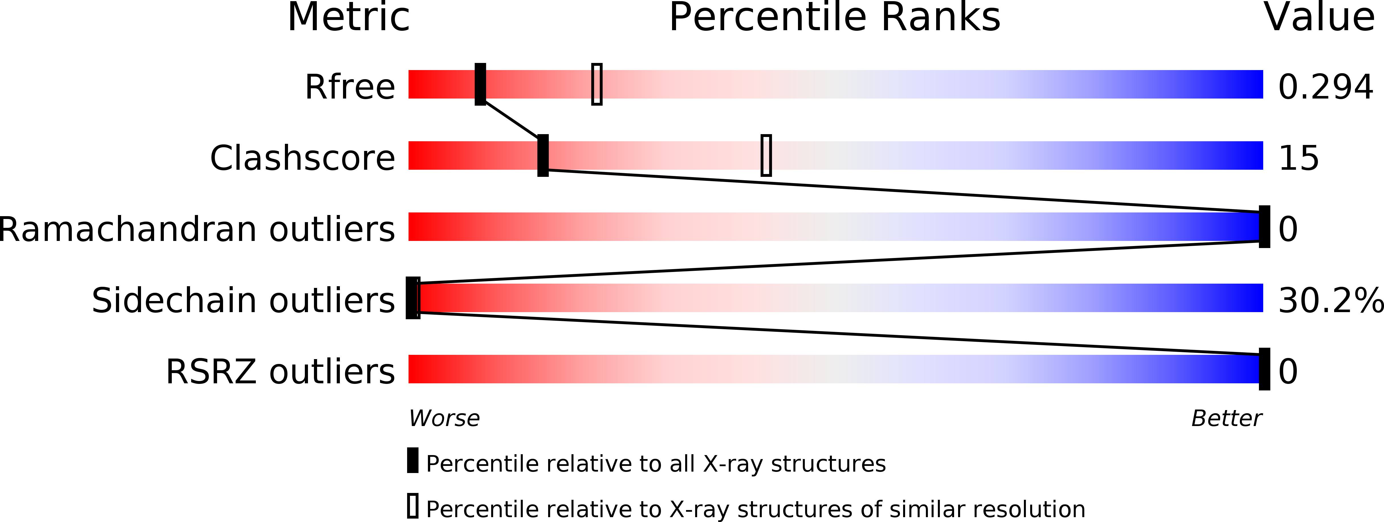
Resolution:
2.80 Å
R-Value Free:
0.30
R-Value Work:
0.27
R-Value Observed:
0.27
Space Group:
P 63
Macromolecular Entities
Polymer Type:polypeptide(L)
Molecule:RHO GUANINE NUCLEOTIDE EXCHANGE FACTOR 7
Gene (Uniprot):Arhgef7
Chain IDs:A
Chain Length:56
Number of Molecules:1
Biological Source:RATTUS NORVEGICUS
Modified Residue
Compound ID Chain ID Parent Comp ID Details 2D Image
MSE A MET SELENOMETHIONINE
Primary Citation
Structures of Dimeric Git1 and Trimeric Beta-Pix and Implications for Git-Pix Complex Assembly.
J.Mol.Biol. 386 280 ? (2009)
PMID: 19136011 DOI: 10.1016/J.JMB.2008.12.050

Abstact

GIT (G protein-coupled receptor kinase-interacting protein) and PIX (p21-activated kinase-interacting exchange factor) family proteins integrate signaling pathways involving Arf and Rho family GTPases. GIT1 and beta-PIX form a constitutively associated complex that acts as a scaffold to allow the formation of large multiprotein assemblies that regulate synaptogenesis, cell polarity and cell migration among other physiological processes. Complex formation is mediated by the GIT binding domain (GBD) in beta-PIX, which recognizes the Spa homology domain of GIT1. Both binding domains are adjacent to predicted coiled-coil segments that allow homo-oligomerization of GIT1 and beta-PIX, respectively. Oligomerization of GIT and PIX proteins is important for their physiological functions, and deletion of the coiled-coil domains interferes with correct subcellular localization and the GEF (guanine nucleotide exchange factor) activity of PIX. We have solved the crystal structures of the CC domains of GIT1 and beta-PIX and determined the stoichiometry of complex formation between the two proteins in order to understand the molecular architecture of the GIT1-beta-PIX complex. The crystal structure of the CC domain of GIT1 solved at 1.4 A resolution shows a dimeric, parallel CC that spans 67 A in length. Unexpectedly, and in contrast to prevalent dimeric models, the structure of the CC region of beta-PIX determined at 2.8 A resolution, combined with hydrodynamic studies, reveals that this protein forms a parallel trimer. Furthermore, we demonstrate that dimeric GIT and trimeric PIX form an unusual high-affinity heteropentameric complex in which each Spa homology domain of the GIT1 dimer recognizes one GBD of the beta-PIX trimer, leaving one GBD unoccupied. These results can serve as a basis to better understand oligomerization-dependent GIT1-beta-PIX-regulated signaling events and provide an insight into the architecture of large signaling complexes involving GIT1 and beta-PIX.

Legend

Protein

Chemical

Disease

Primary Citation of related structures