2W51 image
Deposition Date 2008-12-03
Release Date 2009-03-17
Last Version Date 2024-11-13
Entry Detail
PDB ID:
2W51
Keywords:
Title:
Human mesencephalic astrocyte-derived neurotrophic factor (MANF)
Biological Source:
Source Organism:
HOMO SAPIENS (Taxon ID: 9606)
Host Organism:
Method Details:
Experimental Method:
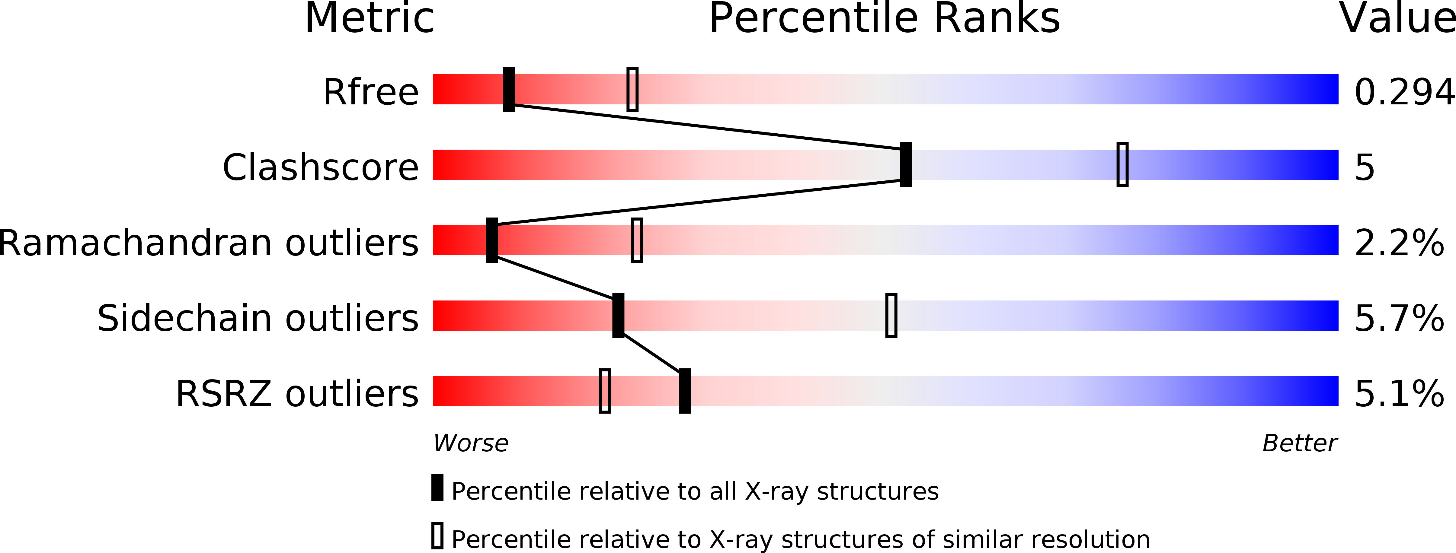
Resolution:
2.80 Å
R-Value Free:
0.30
R-Value Work:
0.28
R-Value Observed:
0.28
Space Group:
P 61
Macromolecular Entities
Structures with similar UniProt ID
Protein Blast
Polymer Type:polypeptide(L)
Molecule:PROTEIN ARMET
Gene (Uniprot):MANF
Chain IDs:A
Chain Length:158
Number of Molecules:1
Biological Source:HOMO SAPIENS
Ligand Molecules
Primary Citation
The Structure of the Conserved Neurotrophic Factors Manf and Cdnf Explains Why They are Bifunctional.
Protein Eng.Des.Sel. 22 233 241 (2009)
PMID: 19258449 DOI: 10.1093/PROTEIN/GZN080

Abstact

We have solved the structures of mammalian mesencephalic astrocyte-derived neurotrophic factor (MANF) and conserved dopamine neurotrophic factor (CDNF). CDNF protects and repairs midbrain dopaminergic neurons in vivo; MANF supports their survival in culture and is also cytoprotective against endoplasmic reticulum (ER) stress. Neither protein structure resembles any known growth factor but the N-terminal domain is a saposin-like lipid-binding domain. MANF and CDNF may thus bind lipids or membranes. Consistent with this, there are two patches of conserved lysines and arginines. The natively unfolded MANF C-terminus contains a CKGC disulphide bridge, such as reductases and disulphide isomerases, consistent with a role in ER stress response. The structure thus explains why MANF and CDNF are bifunctional; neurotrophic activity may reside in the N-terminal domain and ER stress response in the C-terminal domain. Finally, we identified three changes, (MANF)I10-->K(CDNF), (MANF)E79-->M(CDNF) and (MANF)K88-->L(CDNF), that may account for the biological differences between the proteins.

Legend

Protein

Chemical

Disease

Primary Citation of related structures