2W39 image
Deposition Date 2008-11-07
Release Date 2009-07-21
Last Version Date 2024-11-13
Entry Detail
PDB ID:
2W39
Keywords:
Title:
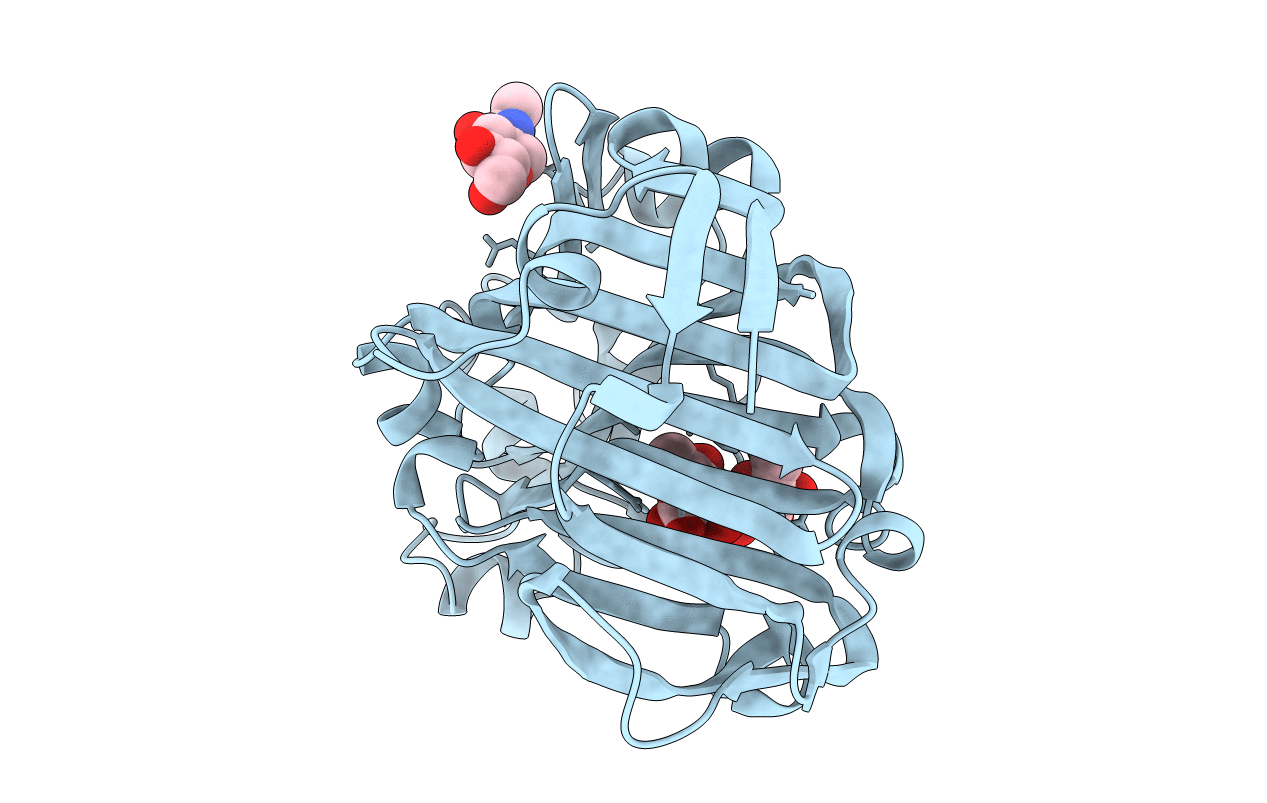
Glc(beta-1-3)Glc disaccharide in -1 and -2 sites of Laminarinase 16A from Phanerochaete chrysosporium
Biological Source:
Source Organism:
Host Organism:
Method Details:
Experimental Method:
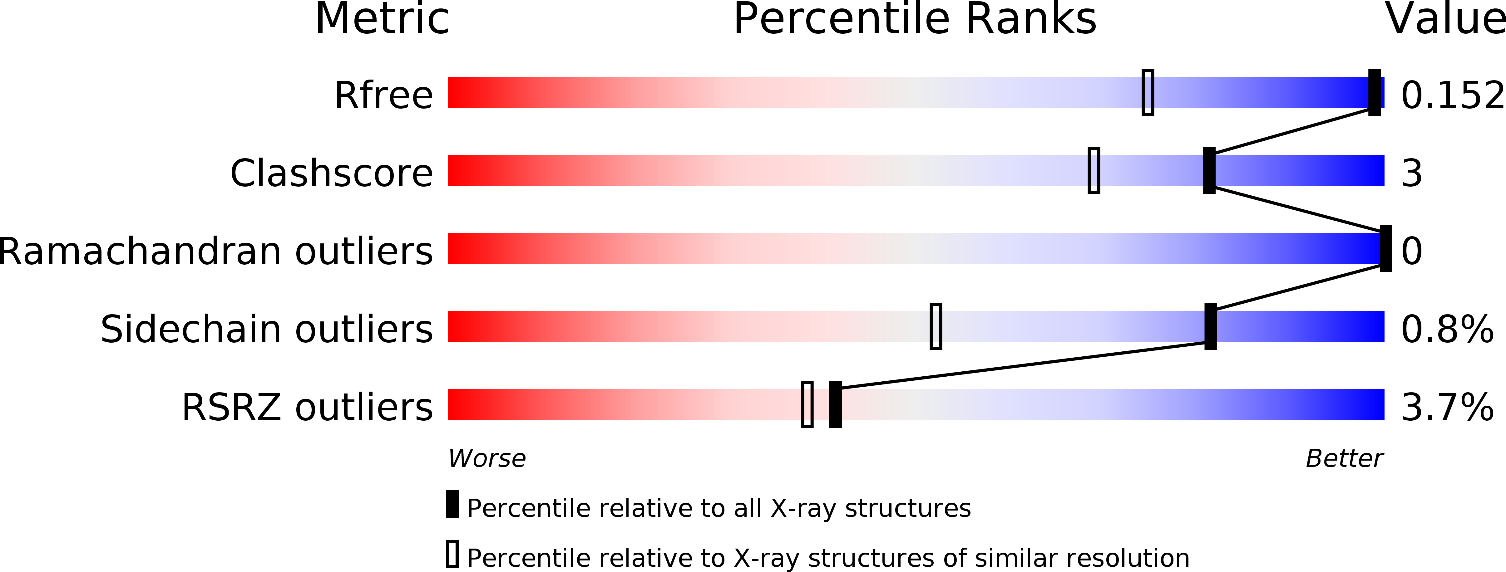
Resolution:
1.10 Å
R-Value Free:
0.15
R-Value Work:
0.13
R-Value Observed:
0.13
Space Group:
P 21 21 21
Macromolecular Entities
Polymer Type:polypeptide(L)
Molecule:PUTATIVE LAMINARINASE
Gene (Uniprot):lam16A
Chain IDs:A
Chain Length:298
Number of Molecules:1
Biological Source:PHANEROCHAETE CHRYSOSPORIUM
Primary Citation
X-Ray Crystal Structures of Phanerochaete Chrysosporium Laminarinase 16A in Complex with Products from Lichenin and Laminarin Hydrolysis
FEBS J. 276 3858 ? (2009)
PMID: 19769746 DOI: 10.1111/J.1742-4658.2009.07099.X

Abstact

The 1,3(4)-beta-D-glucanases of glycoside hydrolase family 16 provide useful examples of versatile yet specific protein-carbohydrate interactions. In the present study, we report the X-ray structures of the 1,3(4)-beta-D-glucanase Phanerochaete chrysosporium Laminarinase 16A in complex with beta-glucan products from laminarin (1.6 A) and lichenin (1.1 A) hydrolysis. The G6G3G3G glucan, in complex with the enzyme, showed a beta-1,6 branch in the acceptor site. The G4G3G ligand-protein complex showed that there was no room for a beta-1,6 branch in the -1 or -2 subsites; furthermore, the distorted residue in the -1 subsite and the glucose in the -2 subsite required a beta-1,3 bond between them. These are the first X-ray crystal structures of any 1,3(4)-beta-D-glucanase in complex with glucan products. They provide details of both substrate and product binding in support of earlier enzymatic evidence.

Legend

Protein

Chemical

Disease

Primary Citation of related structures