Deposition Date
2008-10-30
Release Date
2008-12-09
Last Version Date
2023-12-13
Entry Detail
PDB ID:
2W2H
Keywords:
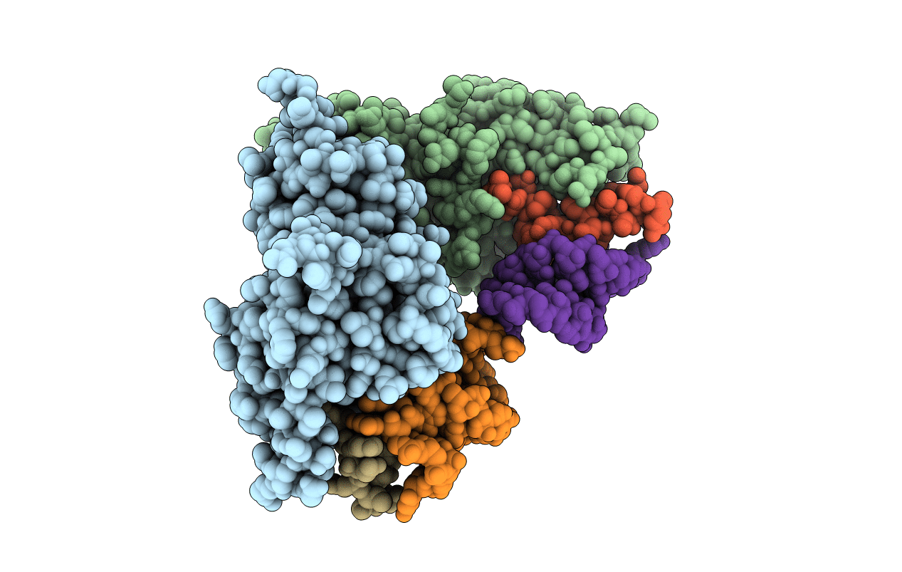
Title:
Structural basis of transcription activation by the Cyclin T1-Tat-TAR RNA complex from EIAV
Biological Source:
Source Organism(s):
EQUUS CABALLUS (Taxon ID: 9796)
EQUINE INFECTIOUS ANEMIA VIRUS (Taxon ID: 11665)
EQUINE INFECTIOUS ANEMIA VIRUS (Taxon ID: 11665)
Expression System(s):
Method Details:
Experimental Method:
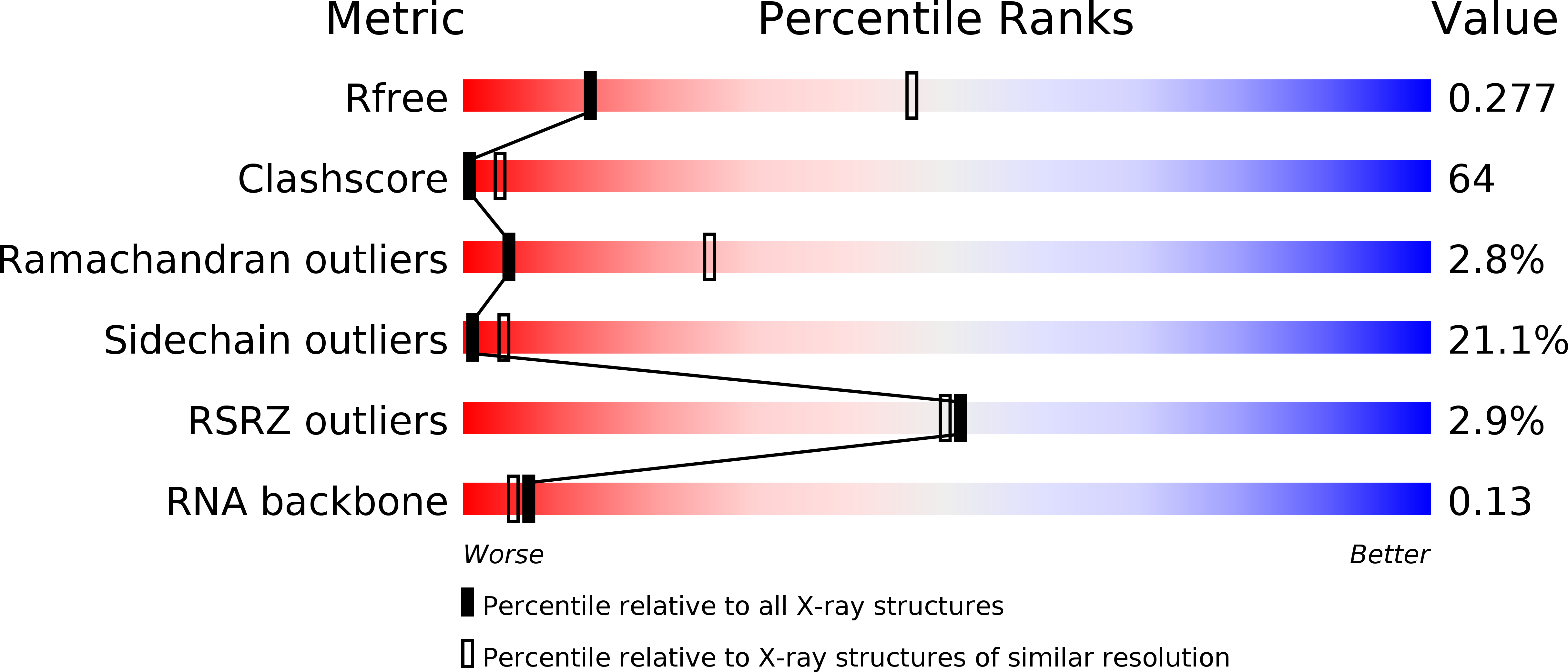
Resolution:
3.25 Å
R-Value Free:
0.27
R-Value Work:
0.24
R-Value Observed:
0.24
Space Group:
I 4