2W07 image
Entry Detail
PDB ID:
2W07
Keywords:
Title:
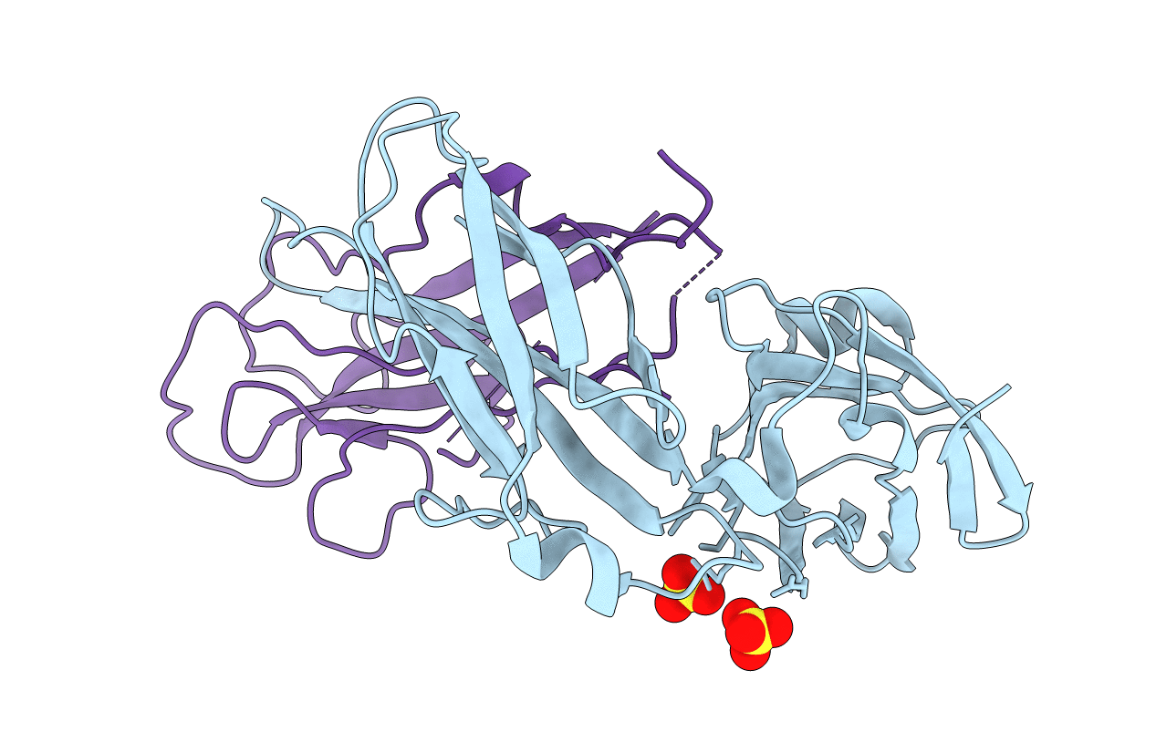
Structural determinants of polymerization reactivity of the P pilus adaptor subunit PapF
Biological Source:
Source Organism:
Host Organism:
PDB Version:
Deposition Date:
2008-08-12
Release Date:
2008-11-25
Method Details:
Experimental Method:
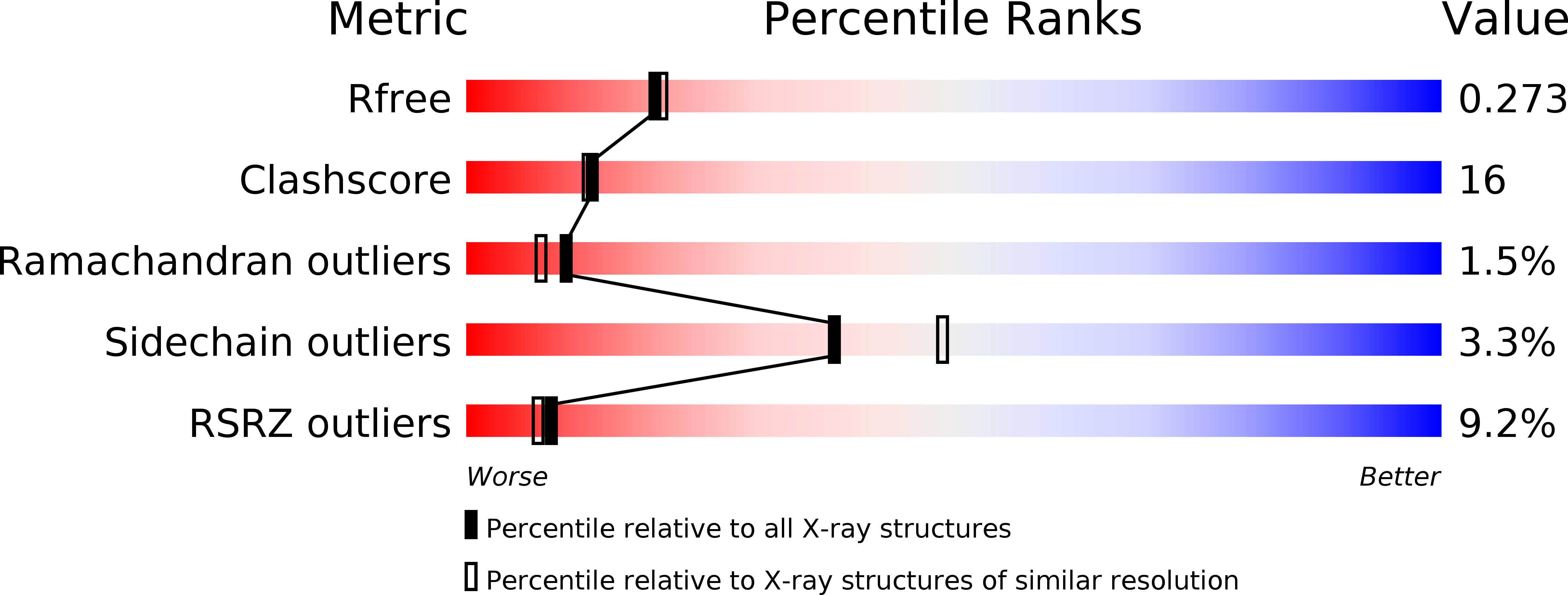
Resolution:
2.20 Å
R-Value Free:
0.27
R-Value Work:
0.23
R-Value Observed:
0.23
Space Group:
P 41 21 2
Macromolecular Entities
Polymer Type:polypeptide(L)
Description:CHAPERONE PROTEIN PAPD
Chain IDs:A
Chain Length:218
Number of Molecules:1
Biological Source:ESCHERICHIA COLI
Polymer Type:polypeptide(L)
Description:MINOR PILIN SUBUNIT PAPF
Mutations:YES
Chain IDs:B
Chain Length:148
Number of Molecules:1
Biological Source:ESCHERICHIA COLI
Ligand Molecules
Primary Citation
Structural Determinants of Polymerization Reactivity of the P Pilus Adaptor Subunit Papf.
Structure 16 1724 ? (2008)
PMID: 19000824 DOI: 10.1016/J.STR.2008.08.012

Abstact

P pili are important adhesive fibers involved in kidney infection by uropathogenic Escherichia coli. Pilus subunits are characterized by a large groove resulting from lack of a beta strand. Polymerization of pilus subunits occurs via the donor-strand exchange (DSE) mechanism initiated when the N terminus of an incoming subunit interacts with the P5 region/pocket of the previously assembled subunit groove. Here, we solve the structure of the PapD:PapF complex in order to understand why PapF undergoes slow DSE. The structure reveals that the PapF P5 pocket is partially obstructed. MD simulations show this region of PapF is flexible compared with its equivalent in PapH, a subunit that also has an obstructed P5 pocket and is unable to undergo DSE. Using electrospray-ionization mass spectrometry, we show that mutations in the P5 region result in increased DSE rates. Thus, partial obstruction of the P5 pocket serves as a modulating mechanism of DSE.

Legend

Protein

Chemical

Disease

Primary Citation of related structures