2VZY image
Deposition Date 2008-08-07
Release Date 2009-04-07
Last Version Date 2024-05-08
Entry Detail
PDB ID:
2VZY
Keywords:
Title:
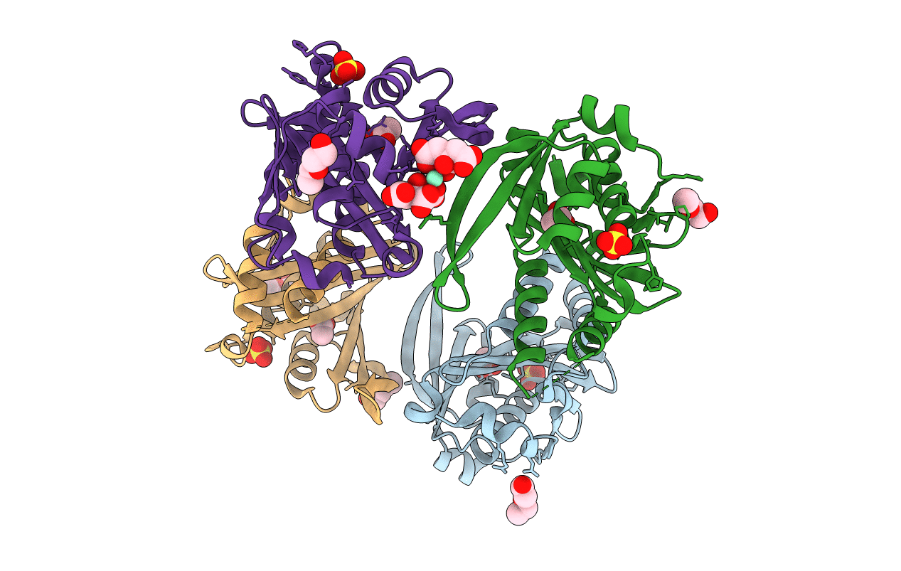
Crystal structure of Rv0802c from Mycobacterium tuberculosis in an unliganded form.
Biological Source:
Source Organism:
Host Organism:
Method Details:
Experimental Method:
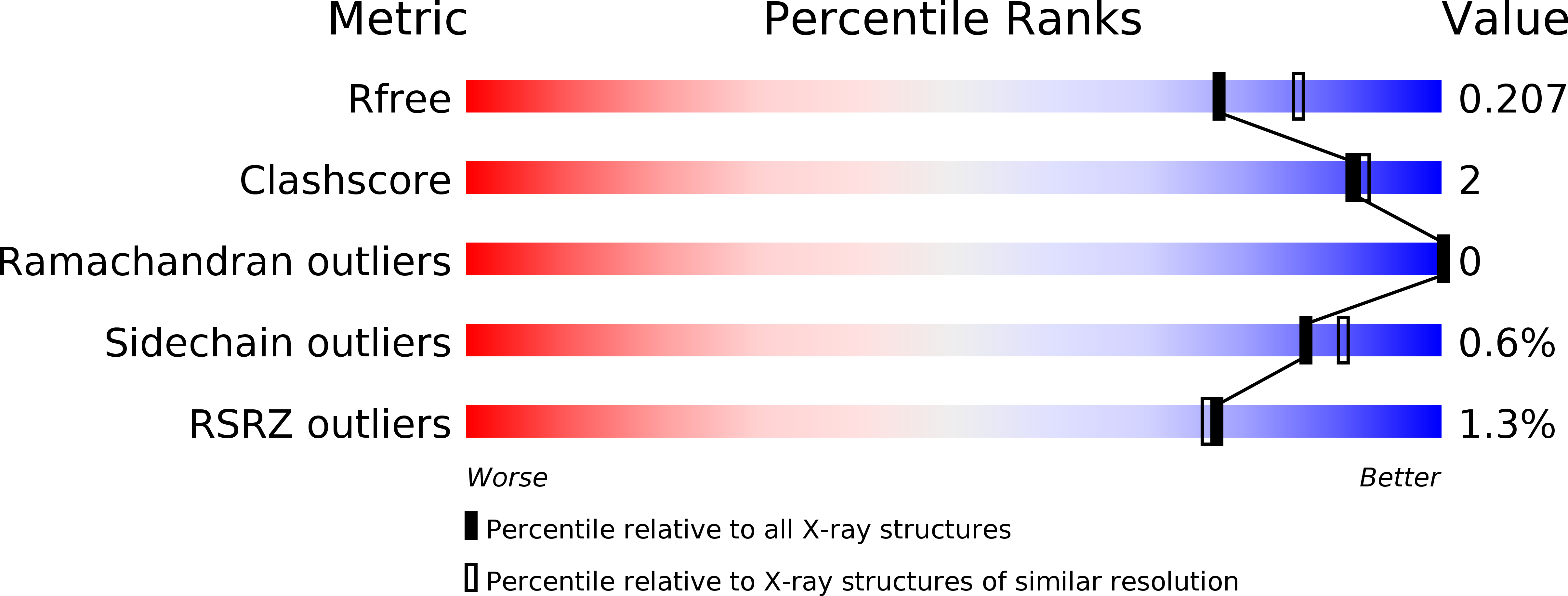
Resolution:
2.00 Å
R-Value Free:
0.20
R-Value Work:
0.16
R-Value Observed:
0.16
Space Group:
P 21 21 21
Macromolecular Entities
Structures with similar UniProt ID
Protein Blast
Polymer Type:polypeptide(L)
Molecule:RV0802C
Chain IDs:A, B, C, D
Chain Length:218
Number of Molecules:4
Biological Source:MYCOBACTERIUM TUBERCULOSIS
Primary Citation
Rv0802C from Mycobacterium Tuberculosis: The First Structure of a Succinyltransferase with the Gnat Fold.
Acta Crystallogr.,Sect.F 64 978 ? (2008)
PMID: 18997321 DOI: 10.1107/S1744309108031679

Abstact

Gene rv0802c from Mycobacterium tuberculosis encodes a 218-amino-acid protein and is annotated as a hypothetical protein with homology to GCN5-related N-acetyltransferases. The structure of Rv0802c was determined in an unliganded form to 2.0 A resolution utilizing single-wavelength anomalous dispersion from a samarium soak that resulted in a single bound Sm(3+):citrate(2) complex. The structure confirms that Rv0802c exhibits the GCN5-related N-acetyltransferase fold and revealed a tetramer composed of a dimer of dimers with approximate 222 symmetry. In addition, a bound acetate ion indicated that Rv0802c may utilize a unique acyl donor for the family. The subsequent determination of the structure of Rv0802c in complex with succinyl-CoA to 2.3 A resolution suggests that Rv0802c is the first known GCN5-related N-acetyltransferase family member to utilize succinyl-CoA as a substrate.

Legend

Protein

Chemical

Disease

Primary Citation of related structures
Feedback Form
Name
Email
Institute
Feedback