2VZC image
Deposition Date 2008-07-31
Release Date 2008-10-28
Last Version Date 2023-12-13
Entry Detail
PDB ID:
2VZC
Keywords:
Title:
Crystal structure of the C-terminal calponin homology domain of alpha parvin
Biological Source:
Source Organism:
HOMO SAPIENS (Taxon ID: 9606)
Host Organism:
Method Details:
Experimental Method:
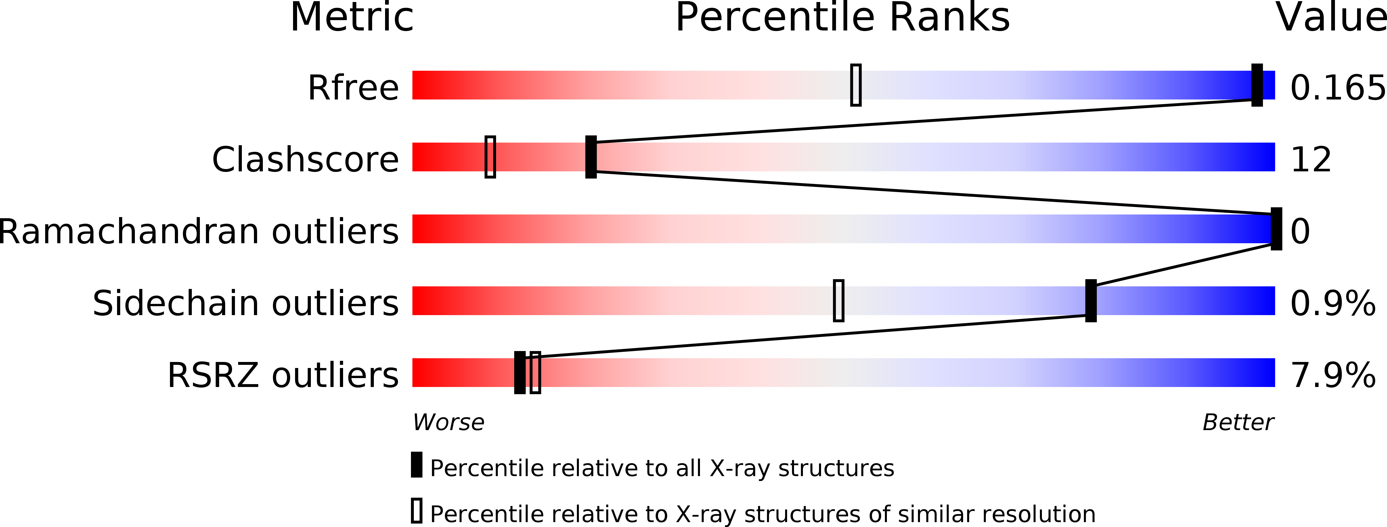
Resolution:
1.05 Å
R-Value Free:
0.15
R-Value Work:
0.14
R-Value Observed:
0.14
Space Group:
P 1 21 1
Macromolecular Entities
Polymer Type:polypeptide(L)
Molecule:ALPHA-PARVIN
Gene (Uniprot):PARVA
Chain IDs:A, B
Chain Length:131
Number of Molecules:2
Biological Source:HOMO SAPIENS
Primary Citation
Structural Analysis of the Interactions between Paxillin Ld Motifs and Alpha-Parvin
Structure 16 1521 ? (2008)
PMID: 18940607 DOI: 10.1016/J.STR.2008.08.007

Abstact

The adaptor protein paxillin contains five conserved leucine-rich (LD) motifs that interact with a variety of focal adhesion proteins, such as alpha-parvin. Here, we report the first crystal structure of the C-terminal calponin homology domain (CH(C)) of alpha-parvin at 1.05 A resolution and show that it is able to bind all the LD motifs, with some selectivity for LD1, LD2, and LD4. Cocrystal structures with these LD motifs reveal the molecular details of their interactions with a common binding site on alpha-parvin-CH(C), which is located at the rim of the canonical fold and includes part of the inter-CH domain linker. Surprisingly, this binding site can accommodate LD motifs in two antiparallel orientations. Taken together, these results reveal an unusual degree of binding degeneracy in the paxillin/alpha-parvin system that may facilitate the assembly of dynamic signaling complexes in the cell.

Legend

Protein

Chemical

Disease

Primary Citation of related structures