2VY0 image
Deposition Date 2008-07-15
Release Date 2009-03-24
Last Version Date 2023-12-13
Entry Detail
PDB ID:
2VY0
Keywords:
Title:
The X-ray structure of endo-beta-1,3-glucanase from Pyrococcus furiosus
Biological Source:
Source Organism:
Host Organism:
Method Details:
Experimental Method:
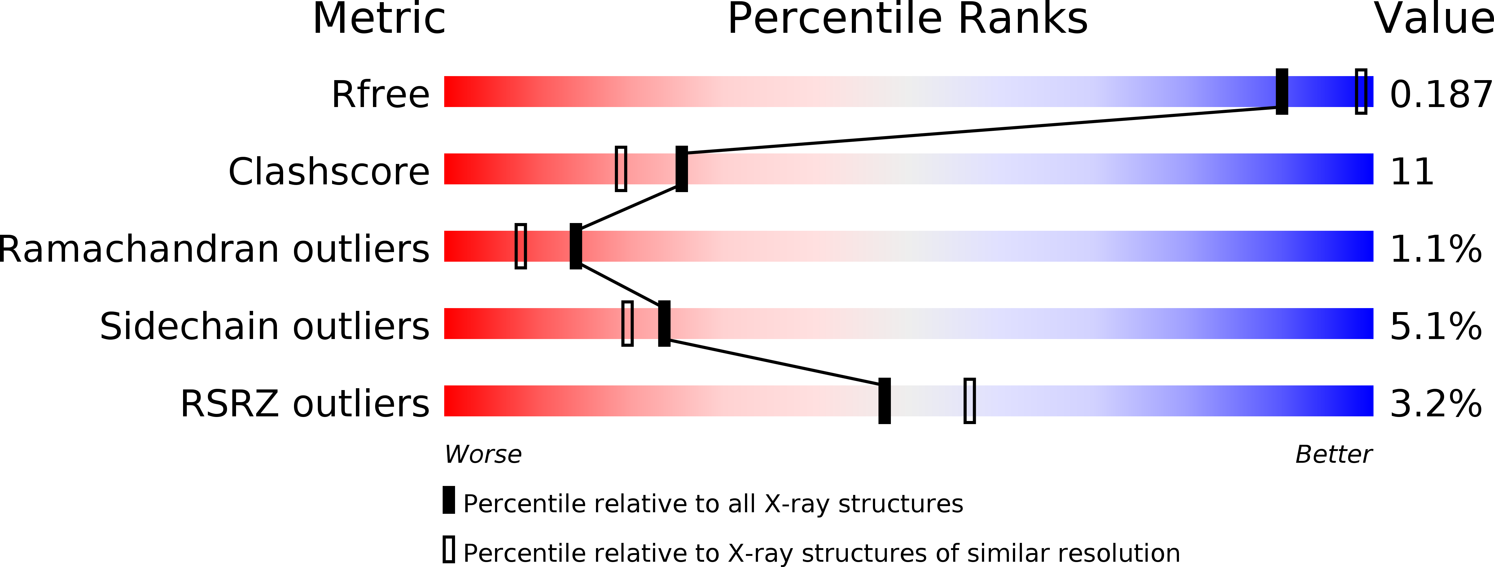
Resolution:
2.16 Å
R-Value Free:
0.22
R-Value Work:
0.19
R-Value Observed:
0.19
Space Group:
P 1 21 1
Macromolecular Entities
Structures with similar UniProt ID
Protein Blast
Polymer Type:polypeptide(L)
Molecule:ENDO-BETA-1,3-GLUCANASE
Gene (Uniprot):lamA
Chain IDs:A, B
Chain Length:264
Number of Molecules:2
Biological Source:PYROCOCCUS FURIOSUS
Primary Citation
Crystal Structure of a Family 16 Endoglucanase from the Hyperthermophile Pyrococcus Furiosus-Structural Basis of Substrate Recognition.
FEBS J. 276 1048 ? (2009)
PMID: 19154353 DOI: 10.1111/J.1742-4658.2008.06848.X

Abstact

Bacterial and archaeal endo-beta-1,3-glucanases that belong to glycoside hydrolase family 16 share a beta-jelly-roll fold, but differ significantly in sequence and in substrate specificity. The crystal structure of the laminarinase (EC 3.2.1.39) from the hyperthermophilic archaeon Pyrococcus furiosus (pfLamA) has been determined at 2.1 A resolution by molecular replacement. The pfLamA structure reveals a kink of six residues (72-77) at the entrance of the catalytic cleft. This peptide is absent in the endoglucanases from alkaliphilic Nocardiopsis sp. strain F96 and Bacillus macerans, two proteins displaying an overall fold similar to that of pfLamA, but with different substrate specificity. A deletion mutant of pfLamA, lacking residues 72-75, hydrolyses the mixed-linkage beta-1,3-1,4-glucan lichenan 10 times more efficiently than the wild-type protein, indicating the importance of the kink in substrate preference.

Legend

Protein

Chemical

Disease

Primary Citation of related structures
Feedback Form
Name
Email
Institute
Feedback