Deposition Date
2008-07-08
Release Date
2009-02-17
Last Version Date
2024-10-16
Entry Detail
PDB ID:
2VXQ
Keywords:
Title:
Crystal structure of the major grass pollen allergen Phl p 2 in complex with its specific IgE-Fab
Biological Source:
Source Organism(s):
PHLEUM PRATENSE (Taxon ID: 15957)
HOMO SAPIENS (Taxon ID: 9606)
HOMO SAPIENS (Taxon ID: 9606)
Expression System(s):
Method Details:
Experimental Method:
Resolution:
1.90 Å
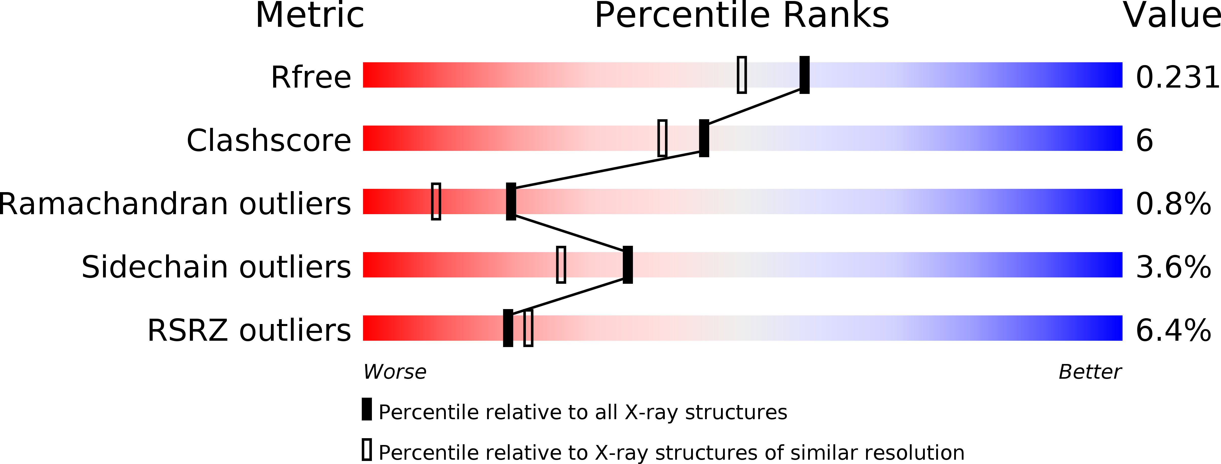
R-Value Free:
0.22
R-Value Work:
0.17
R-Value Observed:
0.17
Space Group:
P 42 21 2