2VXC image
Deposition Date 2008-07-03
Release Date 2008-08-12
Last Version Date 2024-10-09
Entry Detail
PDB ID:
2VXC
Keywords:
Title:
Structure of the Crb2-BRCT2 domain complex with phosphopeptide.
Biological Source:
Source Organism:
Host Organism:
Method Details:
Experimental Method:
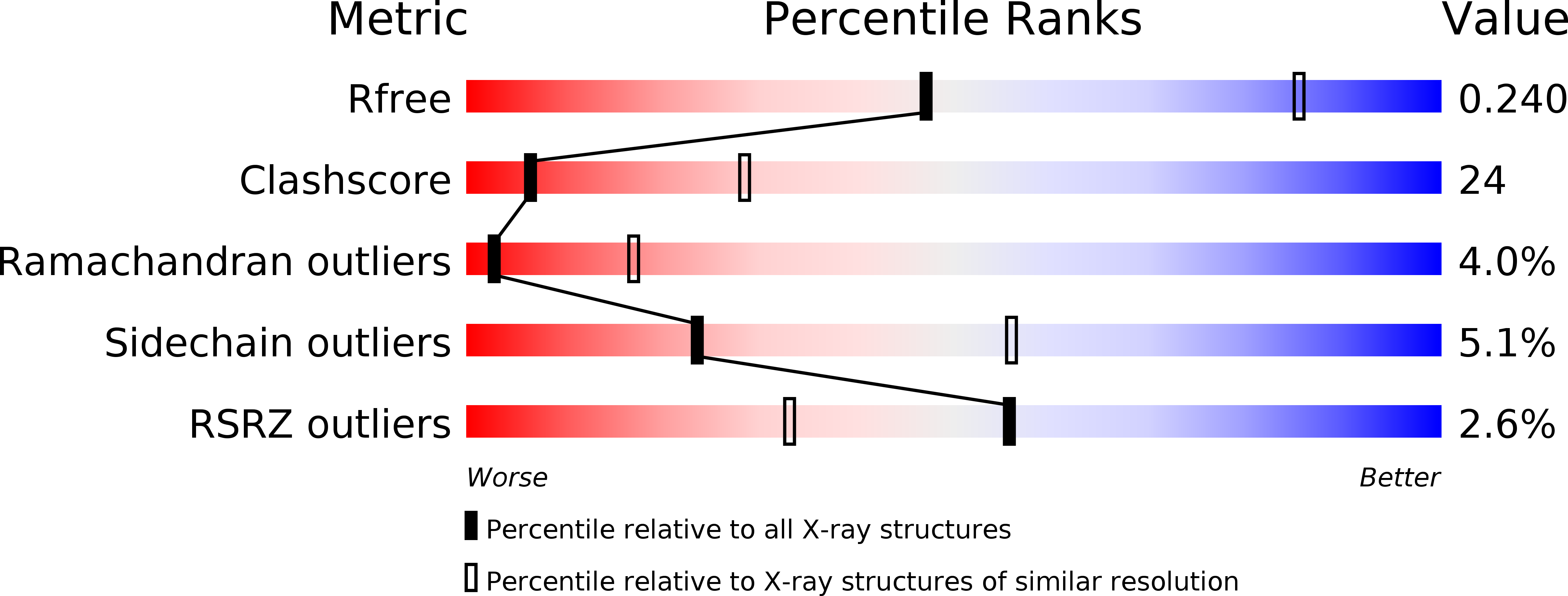
Resolution:
3.10 Å
R-Value Free:
0.24
R-Value Work:
0.19
R-Value Observed:
0.19
Space Group:
P 21 21 2
Macromolecular Entities
Polymer Type:polypeptide(L)
Molecule:DNA REPAIR PROTEIN RHP9
Gene (Uniprot):crb2
Chain IDs:A, B
Chain Length:242
Number of Molecules:2
Biological Source:SCHIZOSACCHAROMYCES POMBE
Polymer Type:polypeptide(L)
Molecule:H2A1 PEPTIDE
Chain IDs:C
Chain Length:4
Number of Molecules:1
Biological Source:HOMO SAPIENS
Modified Residue
Compound ID Chain ID Parent Comp ID Details 2D Image
SEP C SER PHOSPHOSERINE
Ligand Molecules
Primary Citation
Structural and Functional Analysis of the Crb2-Brct2 Domain Reveals Distinct Roles in Checkpoint Signaling and DNA Damage Repair.
Genes Dev. 22 2034 ? (2008)
PMID: 18676809 DOI: 10.1101/GAD.472808

Abstact

Schizosaccharomyces pombe Crb2 is a checkpoint mediator required for the cellular response to DNA damage. Like human 53BP1 and Saccharomyces cerevisiae Rad9 it contains Tudor(2) and BRCT(2) domains. Crb2-Tudor(2) domain interacts with methylated H4K20 and is required for recruitment to DNA dsDNA breaks. The BRCT(2) domain is required for dimerization, but its precise role in DNA damage repair and checkpoint signaling is unclear. The crystal structure of the Crb2-BRCT(2) domain, alone and in complex with a phosphorylated H2A.1 peptide, reveals the structural basis for dimerization and direct interaction with gamma-H2A.1 in ionizing radiation-induced foci (IRIF). Mutational analysis in vitro confirms the functional role of key residues and allows the generation of mutants in which dimerization and phosphopeptide binding are separately disrupted. Phenotypic analysis of these in vivo reveals distinct roles in the DNA damage response. Dimerization mutants are genotoxin sensitive and defective in checkpoint signaling, Chk1 phosphorylation, and Crb2 IRIF formation, while phosphopeptide-binding mutants are only slightly sensitive to IR, have extended checkpoint delays, phosphorylate Chk1, and form Crb2 IRIF. However, disrupting phosphopeptide binding slows formation of ssDNA-binding protein (Rpa1/Rad11) foci and reduces levels of Rad22(Rad52) recombination foci, indicating a DNA repair defect.

Legend

Protein

Chemical

Disease

Primary Citation of related structures