2VW1 image
Deposition Date 2008-06-13
Release Date 2008-06-24
Last Version Date 2023-12-13
Entry Detail
PDB ID:
2VW1
Keywords:
Title:
Crystal structure of the NanB sialidase from Streptococcus pneumoniae
Biological Source:
Source Organism:
Host Organism:
Method Details:
Experimental Method:
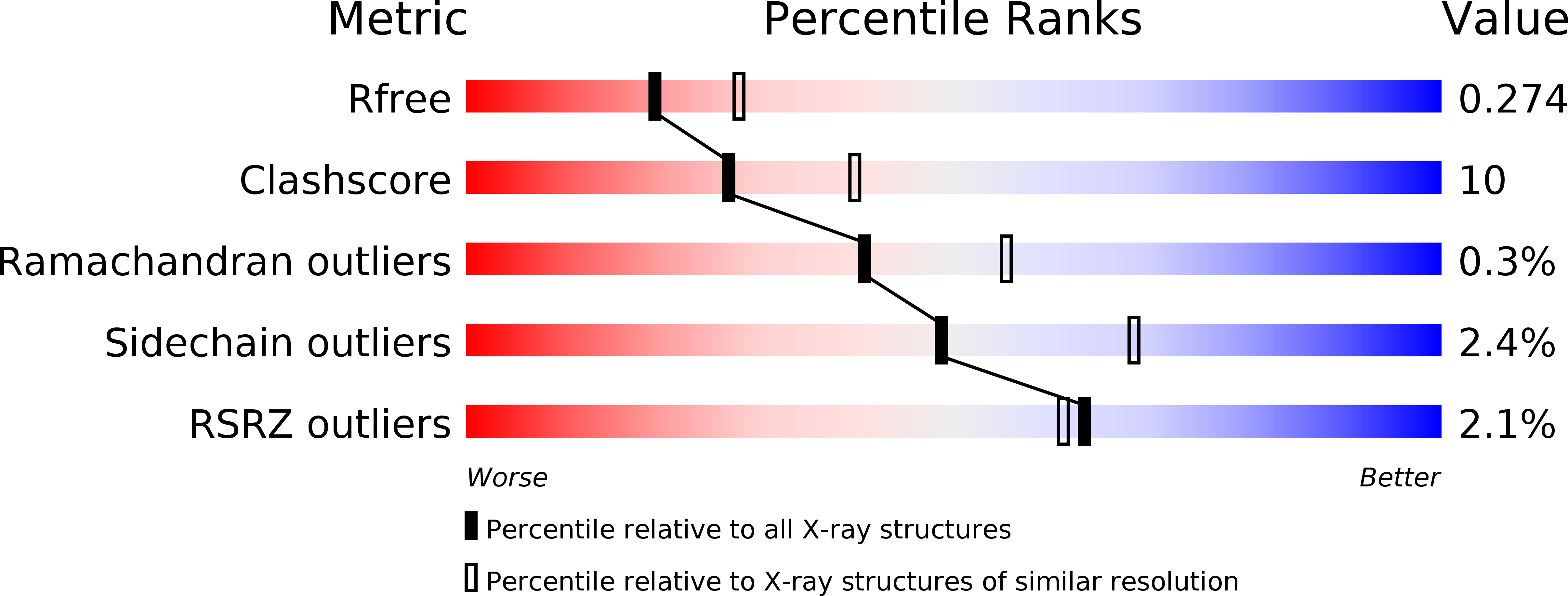
Resolution:
2.39 Å
R-Value Free:
0.27
R-Value Work:
0.19
R-Value Observed:
0.19
Space Group:
P 21 21 21
Macromolecular Entities
Structures with similar UniProt ID
Protein Blast
Polymer Type:polypeptide(L)
Molecule:SIALIDASE B
Gene (Uniprot):nanB
Chain IDs:A
Chain Length:697
Number of Molecules:1
Biological Source:STREPTOCOCCUS PNEUMONIAE
Primary Citation
Crystal Structure of the Nanb Sialidase from Streptococcus Pneumoniae
J.Mol.Biol. 384 436 ? (2008)
PMID: 18835278 DOI: 10.1016/J.JMB.2008.09.032

Abstact

The Streptococcus pneumoniae genomes encode up to three sialidases (or neuraminidases), NanA, NanB and NanC, which are believed to be involved in removing sialic acid from host cell surface glycans, thereby promoting colonization of the upper respiratory tract. Here, we present the crystal structure of NanB to 1.7 A resolution derived from a crystal grown in the presence of the buffer Ches (2-N-cyclohexylaminoethanesulfonic acid). Serendipitously, Ches was found bound to NanB at the enzyme active site, and was found to inhibit NanB with a K(i) of approximately 0.5 mM. In addition, we present the structure to 2.4 A resolution of NanB in complex with the transition-state analogue Neu5Ac2en (2-deoxy-2,3-dehydro-N-acetyl neuraminic acid), which inhibits NanB with a K(i) of approximately 0.3 mM. The sulphonic acid group of Ches and carboxylic acid group of Neu5Ac2en interact with the arginine triad of the active site. The cyclohexyl group of Ches binds in the hydrophobic pocket of NanB occupied by the acetamidomethyl group of Neu5Ac2en. The topology around the NanB active site suggests that the enzyme would have a preference for alpha2,3-linked sialoglycoconjugates, which is confirmed by a kinetic analysis of substrate binding. NMR studies also confirm this preference and show that, like the leech sialidase, NanB acts as an intramolecular trans-sialidase releasing Neu2,7-anhydro5Ac. All three pneumoccocal sialidases possess a carbohydrate-binding domain that is predicted to bind sialic acid. These studies provide support for a possible differential role for NanB compared to NanA in pneumococcal virulence.

Legend

Protein

Chemical

Disease

Primary Citation of related structures