2VVZ image
Deposition Date 2008-06-13
Release Date 2008-06-24
Last Version Date 2023-12-13
Entry Detail
PDB ID:
2VVZ
Keywords:
Title:
Structure of the catalytic domain of Streptococcus pneumoniae sialidase NanA
Biological Source:
Source Organism:
Host Organism:
Method Details:
Experimental Method:
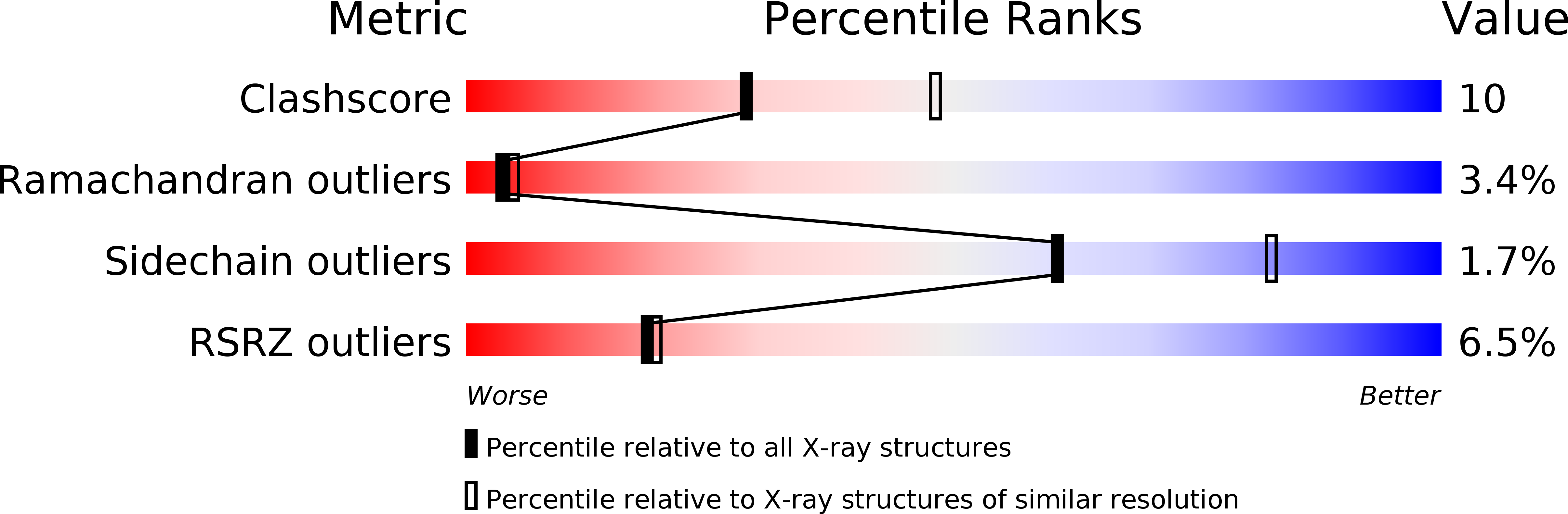
Resolution:
2.50 Å
R-Value Free:
0.29
R-Value Work:
0.24
R-Value Observed:
0.24
Space Group:
P 21 21 21
Macromolecular Entities
Structures with similar UniProt ID
Protein Blast
Polymer Type:polypeptide(L)
Molecule:SIALIDASE A
Gene (Uniprot):nanA
Chain IDs:A, B
Chain Length:504
Number of Molecules:2
Biological Source:STREPTOCOCCUS PNEUMONIAE
Primary Citation
Structure of the Catalytic Domain of Streptococcus Pneumoniae Sialidase Nana.
Acta Crystallogr.,Sect.F 64 772 ? (2008)
PMID: 18765901 DOI: 10.1107/S1744309108024044

Abstact

Streptococcus pneumoniae genomes encode three sialidases, NanA, NanB and NanC, which are key virulence factors that remove sialic acids from various glycoconjugates. The enzymes have potential as drug targets and also as vaccine candidates. The 115 kDa NanA is the largest of the three sialidases and is anchored to the bacterial membrane. Although recombinantly expressed full-length NanA was soluble, it failed to crystallize; therefore, a 56.5 kDa domain that retained full enzyme activity was subcloned. The purified enzyme was crystallized in 0.1 M MES pH 6.5, 30%(w/v) PEG 4000 using the sitting-drop vapour-diffusion method. Data were collected at 100 K to 2.5 A resolution from a crystal grown in the presence of the inhibitor 2-deoxy-2,3-dehydro-N-acetyl neuraminic acid. The crystal belongs to space group P2(1)2(1)2(1), with unit-cell parameters a = 49.2, b = 95.6, c = 226.6 A. The structure was solved by molecular replacement and refined to final R and R(free) factors of 0.246 and 0.298, respectively.

Legend

Protein

Chemical

Disease

Primary Citation of related structures