2VU4 image
Deposition Date 2008-05-21
Release Date 2009-07-21
Last Version Date 2023-12-13
Entry Detail
PDB ID:
2VU4
Keywords:
Title:
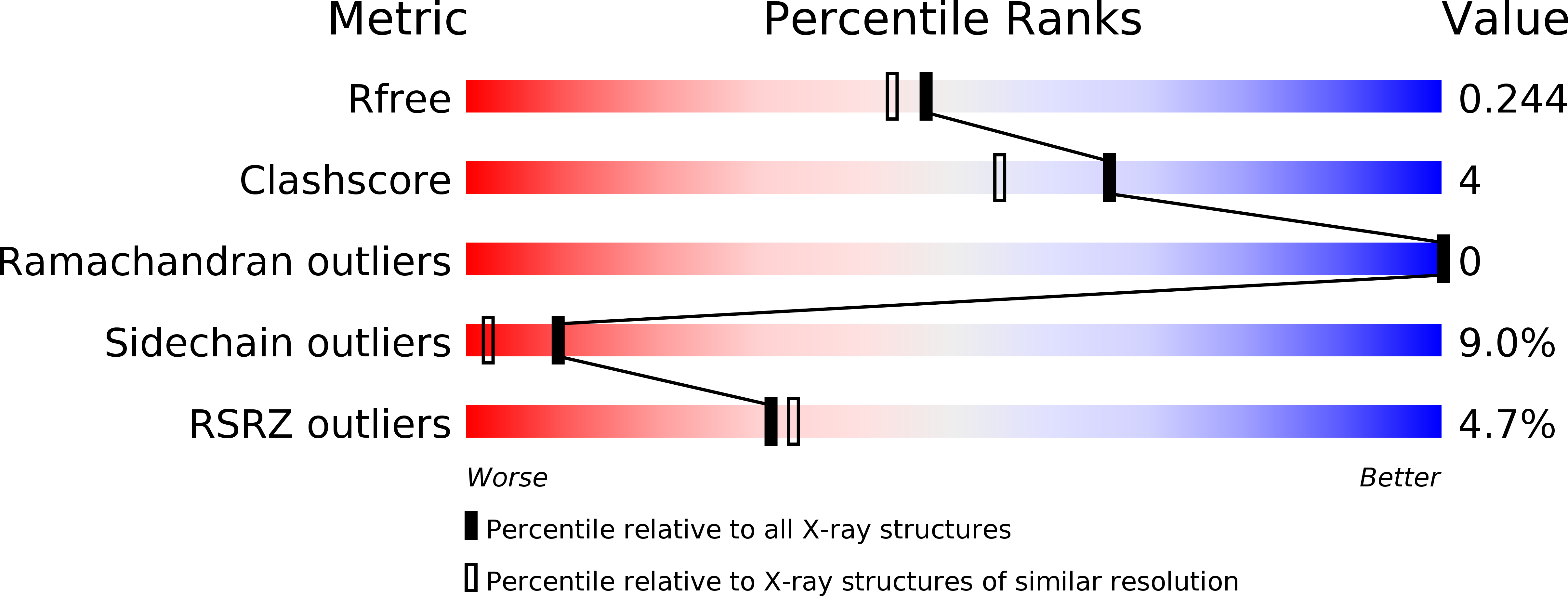
Structure of PsbP protein from Spinacia oleracea at 1.98 A resolution
Biological Source:
Source Organism:
Host Organism:
Method Details:
Experimental Method:
Resolution:
1.98 Å
R-Value Free:
0.23
R-Value Work:
0.18
R-Value Observed:
0.18
Space Group:
P 21 21 21
Macromolecular Entities
Structures with similar UniProt ID
Protein Blast
Polymer Type:polypeptide(L)
Molecule:OXYGEN-EVOLVING ENHANCER PROTEIN 2
Gene (Uniprot):PSBP
Chain IDs:A
Chain Length:190
Number of Molecules:1
Biological Source:SPINACIA OLERACEA
Ligand Molecules
Primary Citation
Raman Spectroscopy Adds Complementary Detail to the High-Resolution X-Ray Crystal Structure of Photosynthetic Psbp from Spinacia Oleracea.
Plos One 7 46694 ? (2012)
PMID: 23071614 DOI: 10.1371/JOURNAL.PONE.0046694

Abstact

Raman microscopy permits structural analysis of protein crystals in situ in hanging drops, allowing for comparison with Raman measurements in solution. Nevertheless, the two methods sometimes reveal subtle differences in structure that are often ascribed to the water layer surrounding the protein. The novel method of drop-coating deposition Raman spectropscopy (DCDR) exploits an intermediate phase that, although nominally "dry," has been shown to preserve protein structural features present in solution. The potential of this new approach to bridge the structural gap between proteins in solution and in crystals is explored here with extrinsic protein PsbP of photosystem II from Spinacia oleracea. In the high-resolution (1.98 Å) x-ray crystal structure of PsbP reported here, several segments of the protein chain are present but unresolved. Analysis of the three kinds of Raman spectra of PsbP suggests that most of the subtle differences can indeed be attributed to the water envelope, which is shown here to have a similar Raman intensity in glassy and crystal states. Using molecular dynamics simulations cross-validated by Raman solution data, two unresolved segments of the PsbP crystal structure were modeled as loops, and the amino terminus was inferred to contain an additional beta segment. The complete PsbP structure was compared with that of the PsbP-like protein CyanoP, which plays a more peripheral role in photosystem II function. The comparison suggests possible interaction surfaces of PsbP with higher-plant photosystem II. This work provides the first complete structural picture of this key protein, and it represents the first systematic comparison of Raman data from solution, glassy, and crystalline states of a protein.

Legend

Protein

Chemical

Disease

Primary Citation of related structures