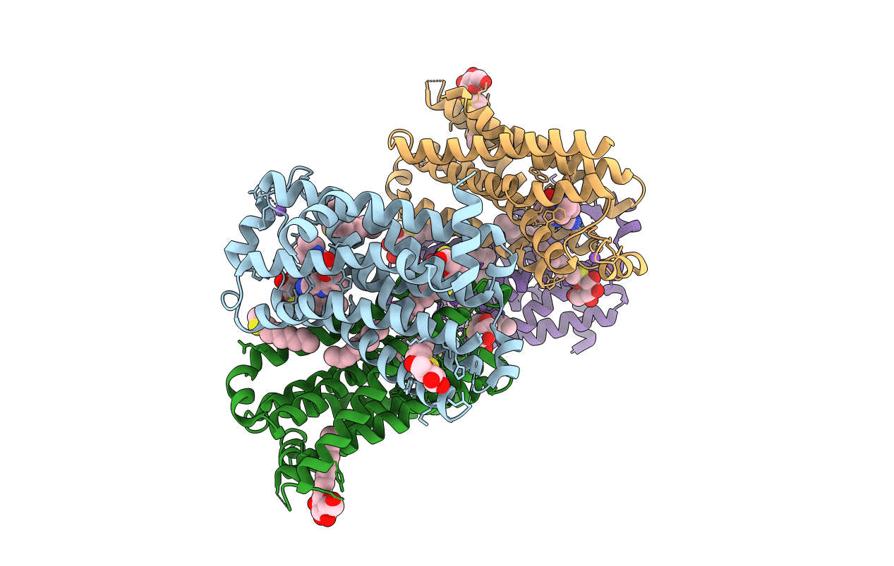
2VT4 image
Deposition Date 2008-05-09
Release Date 2008-06-24
Last Version Date 2025-07-16
Entry Detail
PDB ID:
2VT4
Keywords:
Title:
TURKEY BETA1 ADRENERGIC RECEPTOR WITH STABILISING MUTATIONS AND BOUND CYANOPINDOLOL
Biological Source:
Source Organism:
Host Organism:
Method Details:
Experimental Method:
Resolution:
2.70 Å
R-Value Free:
0.26
R-Value Work:
0.21
R-Value Observed:
0.21
Space Group:
P 1
Macromolecular Entities
Polymer Type:polypeptide(L)
Molecule:BETA1 ADRENERGIC RECEPTOR
Gene (Uniprot):ADRB1
Mutations:YES
Chain IDs:A, B, C, D
Chain Length:313
Number of Molecules:4
Biological Source:MELEAGRIS GALLOPAVO
Primary Citation
Structure of a Beta1-Adrenergic G-Protein-Coupled Receptor.
Nature 454 486 ? (2008)
PMID: 18594507 DOI: 10.1038/NATURE07101

Abstact

G-protein-coupled receptors have a major role in transmembrane signalling in most eukaryotes and many are important drug targets. Here we report the 2.7 A resolution crystal structure of a beta(1)-adrenergic receptor in complex with the high-affinity antagonist cyanopindolol. The modified turkey (Meleagris gallopavo) receptor was selected to be in its antagonist conformation and its thermostability improved by earlier limited mutagenesis. The ligand-binding pocket comprises 15 side chains from amino acid residues in 4 transmembrane alpha-helices and extracellular loop 2. This loop defines the entrance of the ligand-binding pocket and is stabilized by two disulphide bonds and a sodium ion. Binding of cyanopindolol to the beta(1)-adrenergic receptor and binding of carazolol to the beta(2)-adrenergic receptor involve similar interactions. A short well-defined helix in cytoplasmic loop 2, not observed in either rhodopsin or the beta(2)-adrenergic receptor, directly interacts by means of a tyrosine with the highly conserved DRY motif at the end of helix 3 that is essential for receptor activation.

Legend

Protein

Chemical

Disease

Primary Citation of related structures