2VT0 image
Deposition Date 2008-05-03
Release Date 2008-09-23
Last Version Date 2024-10-23
Entry Detail
PDB ID:
2VT0
Keywords:
Title:
X-ray structure of a conjugate with conduritol-beta-epoxide of acid-beta-glucosidase overexpressed in cultured plant cells
Biological Source:
Source Organism:
HOMO SAPIENS (Taxon ID: 9606)
Host Organism:
Method Details:
Experimental Method:
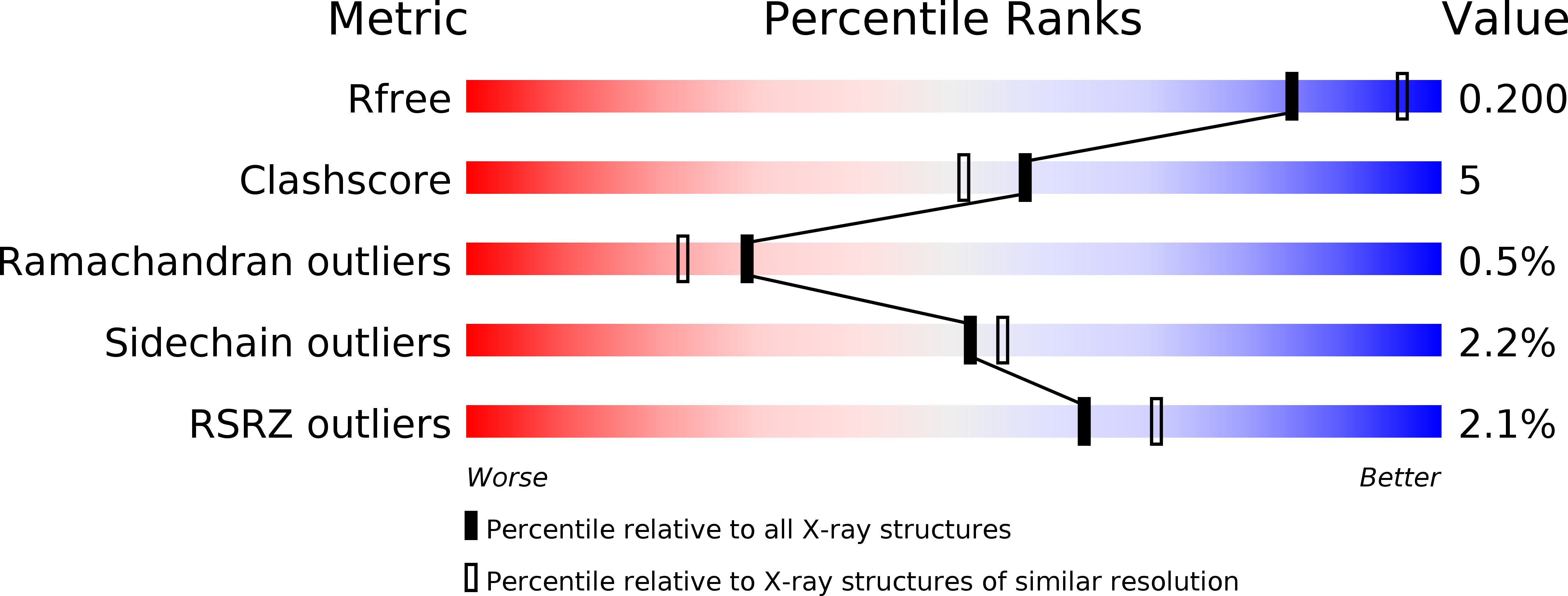
Resolution:
2.15 Å
R-Value Free:
0.20
R-Value Work:
0.15
R-Value Observed:
0.15
Space Group:
P 1 21 1
Macromolecular Entities
Polymer Type:polypeptide(L)
Molecule:GLUCOSYLCERAMIDASE
Chain IDs:A, B
Chain Length:505
Number of Molecules:2
Biological Source:HOMO SAPIENS
Modified Residue
Compound ID Chain ID Parent Comp ID Details 2D Image
ASN A ASN GLYCOSYLATION SITE
Primary Citation
Acid Beta-Glucosidase: Insights from Structural Analysis and Relevance to Gaucher Disease Therapy.
Biol.Chem. 389 1361 ? (2008)
PMID: 18783340 DOI: 10.1515/BC.2008.163

Abstact

In mammalian cells, glucosylceramide (GlcCer), the simplest glycosphingolipid, is hydrolyzed by the lysosomal enzyme acid beta-glucosidase (GlcCerase). In the human metabolic disorder Gaucher disease, GlcCerase activity is significantly decreased owing to one of approximately 200 mutations in the GlcCerase gene. The most common therapy for Gaucher disease is enzyme replacement therapy (ERT), in which patients are given intravenous injections of recombinant human GlcCerase; the Genzyme product Cerezyme has been used clinically for more than 15 years and is administered to approximately 4000 patients worldwide. Here we review the crystal structure of Cerezyme and other recombinant forms of GlcCerase, as well as of their complexes with covalent and non-covalent inhibitors. We also discuss the stability of Cerezyme, which can be altered by modification of its N-glycan chains with possible implications for improved ERT in Gaucher disease.

Legend

Protein

Chemical

Disease

Primary Citation of related structures