Deposition Date
2008-04-29
Release Date
2008-05-27
Last Version Date
2023-12-13
Entry Detail
PDB ID:
2VSS
Keywords:
Title:
Wild-type Hydroxycinnamoyl-CoA hydratase lyase in complex with acetyl- CoA and vanillin
Biological Source:
Source Organism(s):
PSEUDOMONAS FLUORESCENS (Taxon ID: 294)
Expression System(s):
Method Details:
Experimental Method:
Resolution:
2.22 Å
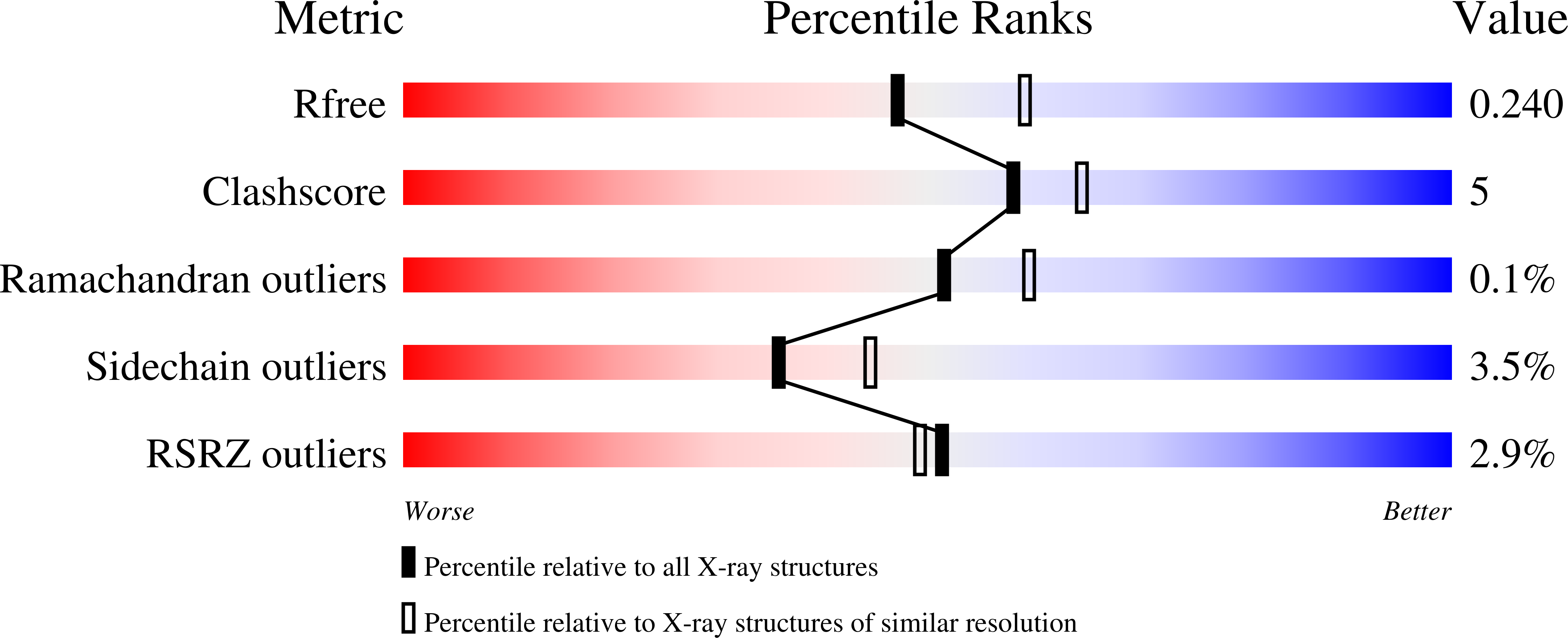
R-Value Free:
0.24
R-Value Work:
0.18
R-Value Observed:
0.18
Space Group:
P 21 21 21