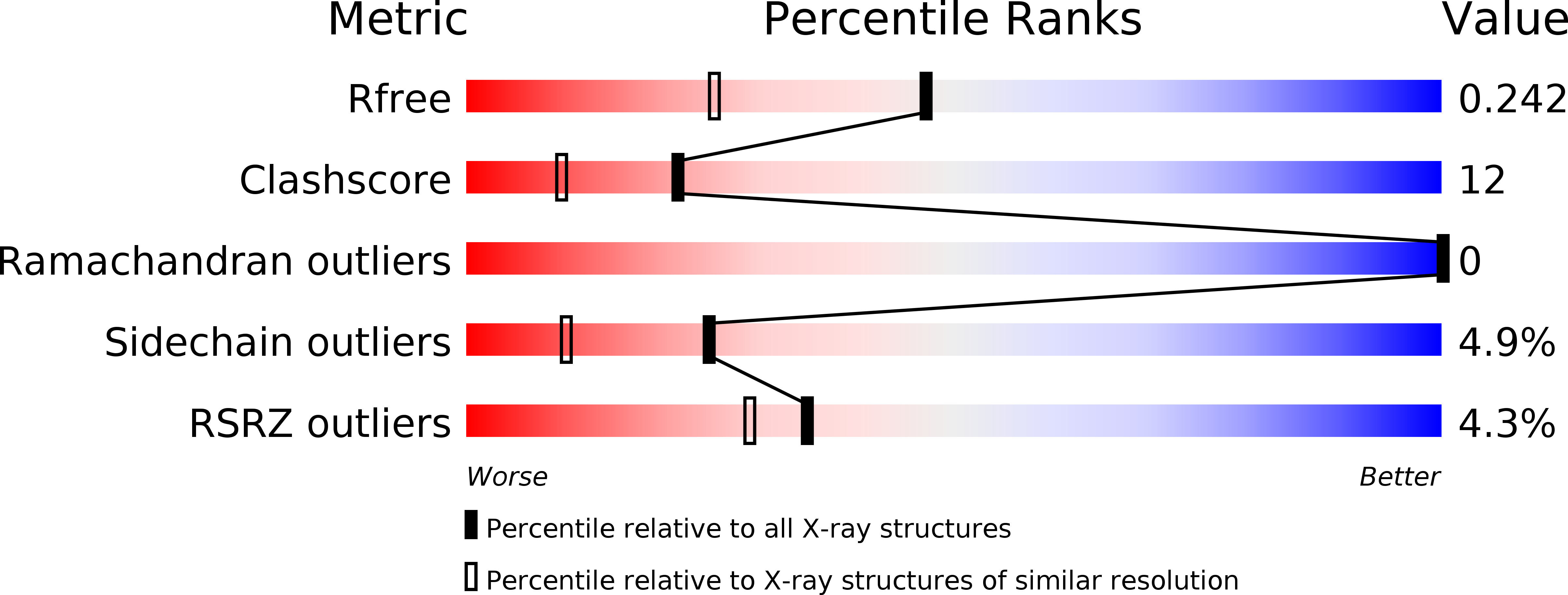
2VSD image
Entry Detail
PDB ID:
2VSD
Title:
crystal structure of CHIR-AB1
Biological Source:
Source Organism:
Host Organism:
PDB Version:
Deposition Date:
2008-04-22
Release Date:
2008-07-29
Method Details:
Experimental Method:
Resolution:
1.82 Å
R-Value Free:
0.24
R-Value Work:
0.20
R-Value Observed:
0.21
Space Group:
P 31 2 1
Macromolecular Entities
Polymer Type:polypeptide(L)
Description:CHIR AB1
Chain IDs:A
Chain Length:105
Number of Molecules:1
Biological Source:GALLUS GALLUS
Modified Residue
Compound ID Chain ID Parent Comp ID Details 2D Image
ASN A ASN GLYCOSYLATION SITE
Primary Citation
The Crystal Structure of Chir-Ab1: A Primordial Avian Classical Fc Receptor.
J.Mol.Biol. 381 1012 ? (2008)
PMID: 18625238 DOI: 10.1016/J.JMB.2008.06.082

Abstact

CHIR-AB1 is a newly identified avian immunoglobulin (Ig) receptor that includes both activating and inhibitory motifs and was therefore classified as a potentially bifunctional receptor. Recently, CHIR-AB1 was shown to bind the Fc region of chicken IgY and to induce calcium mobilization via association with the common gamma-chain, a subunit that transmits signals upon ligation of many different immunoreceptors. Here we describe the 1.8-A-resolution crystal structure of the CHIR-AB1 ectodomain. The receptor ectodomain consists of a single C2-type Ig domain resembling the Ig-like domains found in mammalian Fc receptors such as FcgammaRs and FcalphaRI. Unlike these receptors and other monomeric Ig superfamily members, CHIR-AB1 crystallized as a 2-fold symmetrical homodimer that bears no resemblance to variable or constant region dimers in an antibody. Analytical ultracentrifugation demonstrated that CHIR-AB1 exists as a mixture of monomers and dimers in solution, and equilibrium gel filtration revealed a 2:1 receptor/ligand binding stoichiometry. Measurement of the 1:1 CHIR-AB1/IgY interaction affinity indicates a relatively low affinity complex, but a 2:1 CHIR-AB1/IgY interaction allows an increase in apparent affinity due to avidity effects when the receptor is tethered to a surface. Taken together, these results add to the structural understanding of Fc receptors and their functional mechanisms.

Legend

Protein

Chemical

Disease

Primary Citation of related structures