2VRK image
Entry Detail
PDB ID:
2VRK
Keywords:
Title:
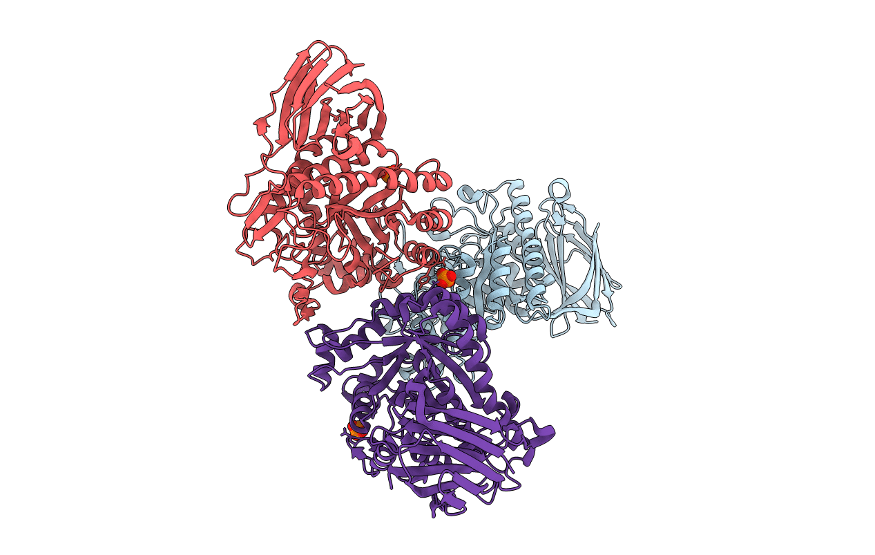
Structure of a seleno-methionyl derivative of wild type arabinofuranosidase from Thermobacillus xylanilyticus
Biological Source:
Host Organism:
PDB Version:
Deposition Date:
2008-04-09
Release Date:
2008-07-01
Method Details:
Experimental Method:
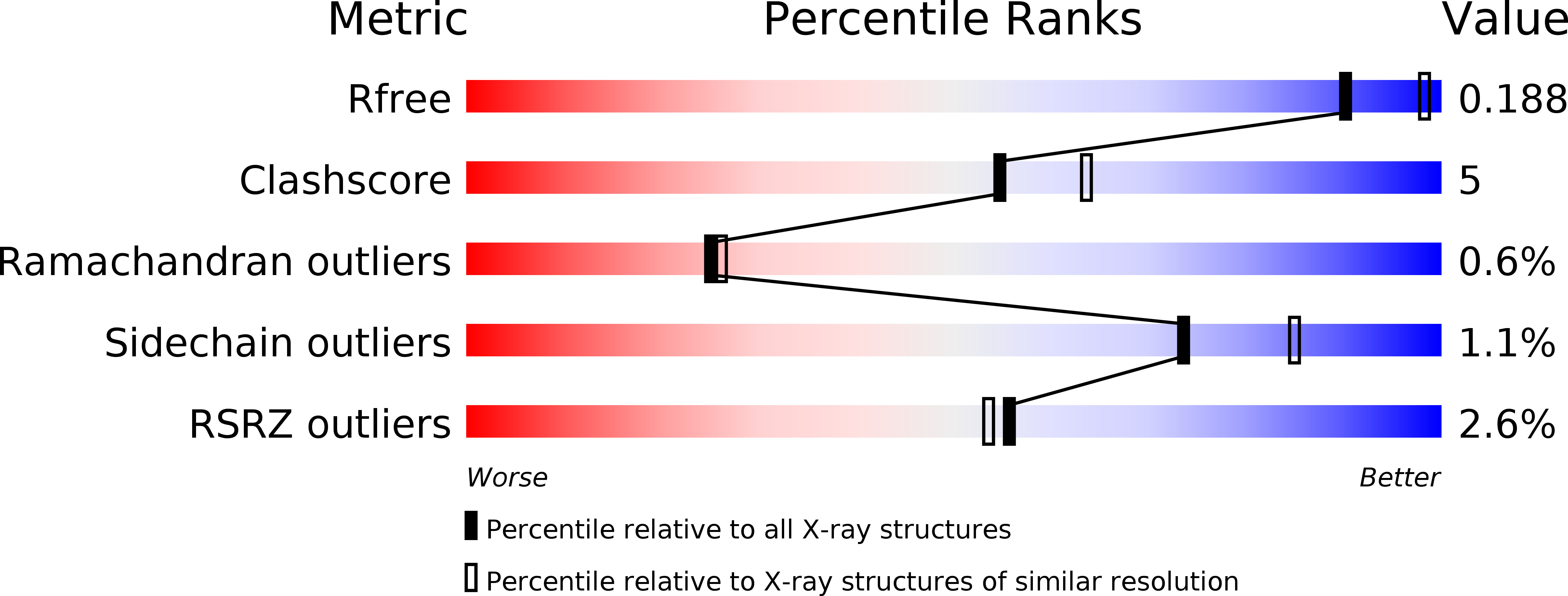
Resolution:
2.20 Å
R-Value Free:
0.18
R-Value Work:
0.16
R-Value Observed:
0.16
Space Group:
P 65 2 2
Macromolecular Entities
Polymer Type:polypeptide(L)
Description:ALPHA-L-ARABINOFURANOSIDASE
Chain IDs:A, B, C
Chain Length:496
Number of Molecules:3
Biological Source:THERMOBACILLUS XYLANILYTICUS
Modified Residue
Compound ID Chain ID Parent Comp ID Details 2D Image
MSE A MET SELENOMETHIONINE
Ligand Molecules
Primary Citation
The Structure of the Complex between a Branched Pentasaccharide and Thermobacillus Xylanilyticus Gh-51 Arabinofuranosidase Reveals Xylan-Binding Determinants and Induced Fit.
Biochemistry 47 7441 ? (2008)
PMID: 18563919 DOI: 10.1021/BI800424E

Abstact

The crystal structure of the family GH-51 alpha- l-arabinofuranosidase from Thermobacillus xylanilyticus has been solved as a seleno-methionyl derivative. In addition, the structure of an inactive mutant Glu176Gln is presented in complex with a branched pentasaccharide, a fragment of its natural substrate xylan. The overall structure shows the two characteristic GH-51 domains: a catalytic domain that is folded into a (beta/alpha) 8-barrel and a C-terminal domain that displays jelly roll architecture. The pentasaccharide is bound in a groove on the surface of the enzyme, with the mono arabinosyl branch entering a tight pocket harboring the catalytic dyad. Detailed analyses of both structures and comparisons with the two previously determined structures from Geobacillus stearothermophilus and Clostridium thermocellum reveal important details unique to the Thermobacillus xylanilyticus enzyme. In the absence of substrate, the enzyme adopts an open conformation. In the substrate-bound form, the long loop connecting beta-strand 2 to alpha-helix 2 closes the active site and interacts with the substrate through residues His98 and Trp99. The results of kinetic and fluorescence titration studies using mutants underline the importance of this loop, and support the notion of an interaction between Trp99 and the bound substrate. We suggest that the changes in loop conformation are an integral part of the T. xylanilyticus alpha- l-arabinofuranosidase reaction mechanism, and ensure efficient binding and release of substrate.

Legend

Protein

Chemical

Disease

Primary Citation of related structures