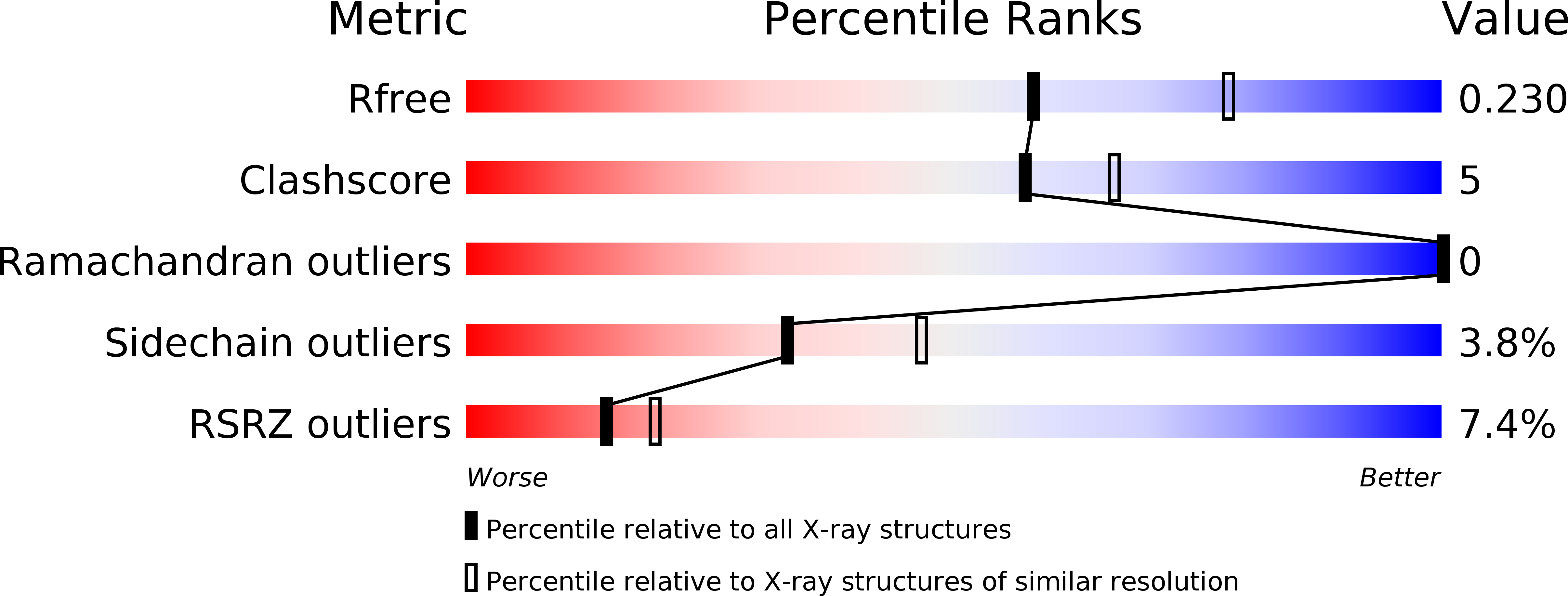
2VQZ image
Deposition Date 2008-03-21
Release Date 2008-05-13
Last Version Date 2024-10-23
Entry Detail
PDB ID:
2VQZ
Keywords:
Title:
Structure of the cap-binding domain of influenza virus polymerase subunit PB2 with bound m7GTP
Biological Source:
Source Organism:
Host Organism:
Method Details:
Experimental Method:
Resolution:
2.30 Å
R-Value Free:
0.23
R-Value Work:
0.18
R-Value Observed:
0.18
Space Group:
C 2 2 21
Macromolecular Entities
Polymer Type:polypeptide(L)
Molecule:POLYMERASE BASIC PROTEIN 2
Gene (Uniprot):PB2
Mutations:YES
Chain IDs:A, B, C (auth: D), D (auth: E), E (auth: F)
Chain Length:166
Number of Molecules:5
Biological Source:INFLUENZA A VIRUS
Modified Residue
Compound ID Chain ID Parent Comp ID Details 2D Image
MSE A MET SELENOMETHIONINE
Ligand Molecules
Primary Citation
The Structural Basis for CAP Binding by Influenza Virus Polymerase Subunit Pb2.
Nat.Struct.Mol.Biol. 15 500 ? (2008)
PMID: 18454157 DOI: 10.1038/NSMB.1421

Abstact

Influenza virus mRNAs are synthesized by the trimeric viral polymerase using short capped primers obtained by a 'cap-snatching' mechanism. The polymerase PB2 subunit binds the 5' cap of host pre-mRNAs, which are cleaved after 10-13 nucleotides by the PB1 subunit. Using a library-screening method, we identified an independently folded domain of PB2 that has specific cap binding activity. The X-ray structure of the domain with bound cap analog m(7)GTP at 2.3-A resolution reveals a previously unknown fold and a mode of ligand binding that is similar to, but distinct from, other cap binding proteins. Binding and functional studies with point mutants confirm that the identified site is essential for cap binding in vitro and cap-dependent transcription in vivo by the trimeric polymerase complex. These findings clarify the nature of the cap binding site in PB2 and will allow efficient structure-based design of new anti-influenza compounds inhibiting viral transcription.

Legend

Protein

Chemical

Disease

Primary Citation of related structures