Deposition Date
2008-03-18
Release Date
2009-02-17
Last Version Date
2024-05-08
Entry Detail
PDB ID:
2VQP
Keywords:
Title:
Structure of the matrix protein from human Respiratory Syncytial Virus
Biological Source:
Source Organism(s):
HUMAN RESPIRATORY SYNCYTIAL VIRUS (Taxon ID: 11259)
Expression System(s):
Method Details:
Experimental Method:
Resolution:
1.60 Å
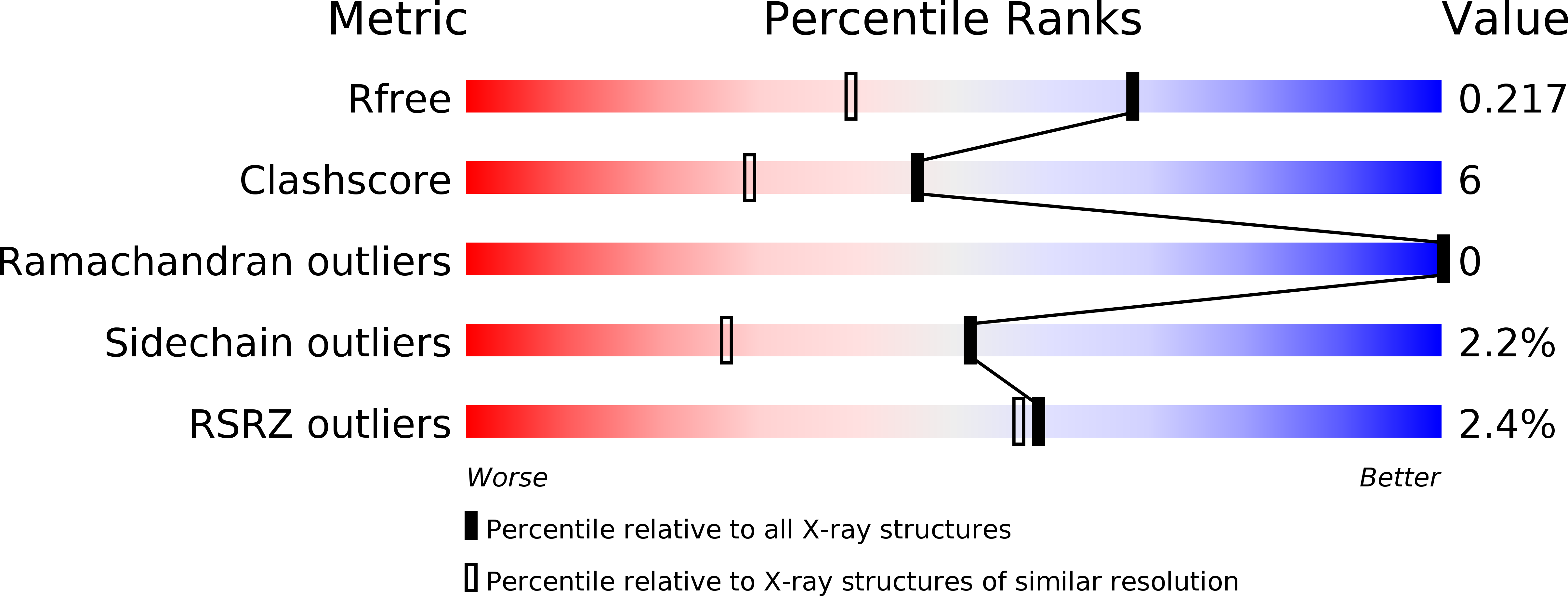
R-Value Free:
0.21
R-Value Work:
0.18
R-Value Observed:
0.18
Space Group:
C 1 2 1