2VPL image
Deposition Date 2008-03-01
Release Date 2008-09-23
Last Version Date 2023-12-13
Entry Detail
PDB ID:
2VPL
Keywords:
Title:
The structure of the complex between the first domain of L1 protein from Thermus thermophilus and mRNA from Methanococcus jannaschii
Biological Source:
Source Organism:
Host Organism:
Method Details:
Experimental Method:
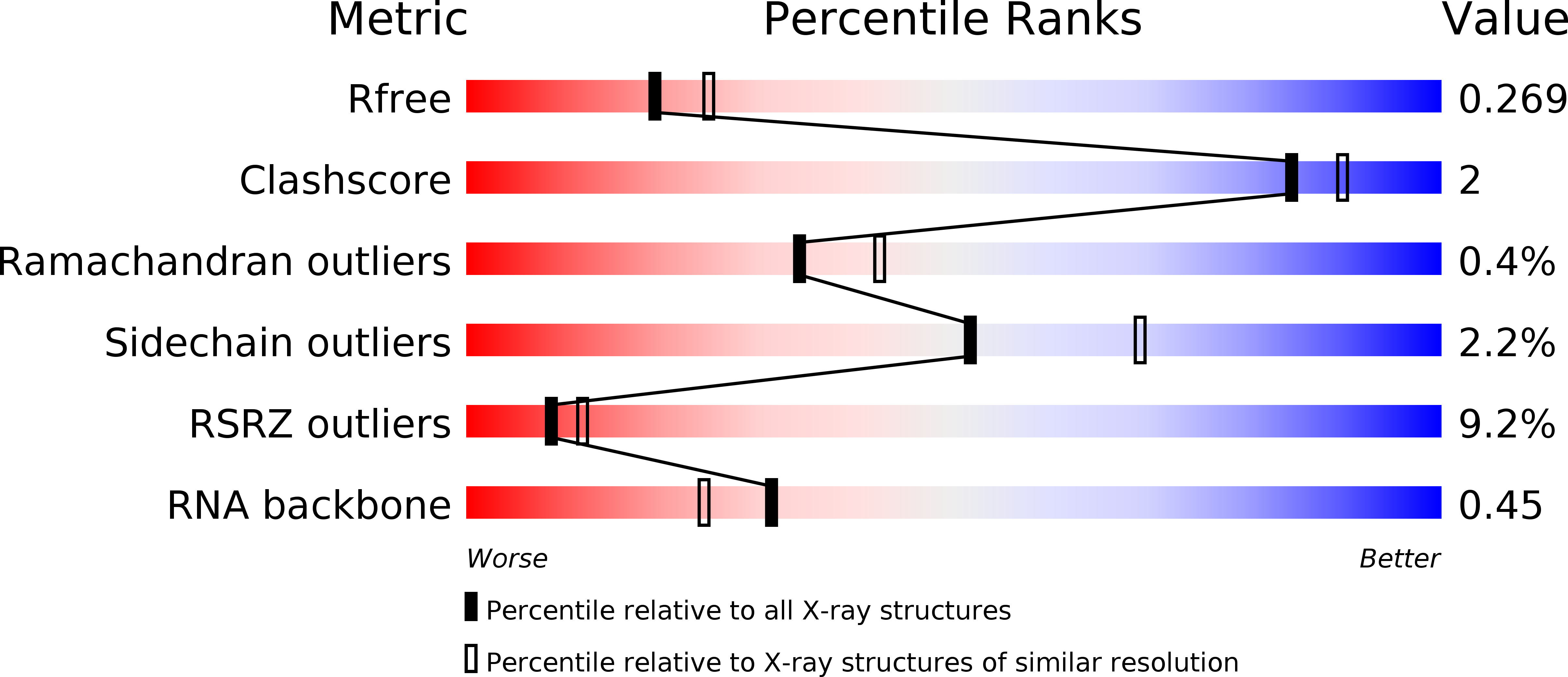
Resolution:
2.30 Å
R-Value Free:
0.27
R-Value Work:
0.20
R-Value Observed:
0.21
Space Group:
P 21 21 2
Macromolecular Entities
Structures with similar UniProt ID
Protein Blast
Polymer Type:polypeptide(L)
Molecule:50S RIBOSOMAL PROTEIN L1
Gene (Uniprot):rplA
Chain IDs:A, C
Chain Length:137
Number of Molecules:2
Biological Source:THERMUS THERMOPHILUS
Polymer Type:polyribonucleotide
Molecule:FRAGMENT OF MRNA FOR L1-OPERON CONTAINING REGULATOR L1-BINDING SITE
Chain IDs:B, D
Chain Length:48
Number of Molecules:2
Biological Source:METHANOCALDOCOCCUS JANNASCHII
Primary Citation
Domain II of Thermus thermophilus ribosomal protein L1 hinders recognition of its mRNA.
J. Mol. Biol. 383 301 305 (2008)
PMID: 18778715 DOI: 10.1016/j.jmb.2008.08.058

Abstact

The two-domain ribosomal protein L1 has a dual function as a primary rRNA-binding ribosomal protein and as a translational repressor that binds its own mRNA. Here, we report the crystal structure of a complex between the isolated domain I of L1 from the bacterium Thermus thermophilus and a specific mRNA fragment from Methanoccocus vannielii. In parallel, we report kinetic characteristics measured for complexes formed by intact TthL1 and its domain I with the specific mRNA fragment. Although, there is a close similarity between the RNA-protein contact regions in both complexes, the association rate constant is higher in the case of the complex formed by the isolated domain I. This finding demonstrates that domain II hinders mRNA recognition by the intact TthL1.

Legend

Protein

Chemical

Disease

Primary Citation of related structures